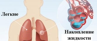
Pulmonary emphysema is a persistent increase in lung space, which is accompanied by destruction of the walls of the alveoli and other structural elements without the proliferation of connective tissue. The physiological feature of the bronchi is expansion on inhalation and slight narrowing on exhalation. With emphysema, a person takes a fairly effective breath, but cannot fully exhale. This leads to the accumulation of air in the lung tissues, while the alveoli swell and burst, and large air sacs (bullas) appear. Gas exchange worsens, the lungs become flabby and do not collapse. This is how pulmonary emphysema develops, the treatment of which is limited, since the changes occurring in the lungs are irreversible. Patients are generally recommended to reduce physical activity and avoid smoking. In case of severe respiratory failure, the patient is connected to portable oxygen therapy devices.
Types of emphysema
The classification of emphysema is as follows.
Forms of the disease according to the course:
- Acute - develops with sudden stress, penetration of foreign bodies into the lungs, asthma. This condition can be eliminated, but requires immediate medical intervention.
- Chronic - progresses gradually, leading to disability without taking action.
By etiology:
- Primary - develops due to the individual characteristics of the body, can be observed even in newborns. It is characterized by rapid progression and complex therapy.
- Secondary - develops against the background of obstruction of lung tissue.
By degree of distribution:
- Diffuse - CT shows that the tissue is affected evenly, destruction of the alveoli occurs throughout the entire area, and the patient is bothered by shortness of breath.
- Focal - changes are localized near scars, foci of tuberculosis. This is clearly visible on x-rays.
According to anatomical features:
- Panacinar - all acini are damaged, there is no proliferation of connective tissue and no inflammation, but there are signs of impaired gas exchange in the lungs (the type of breathing resembles panting).
- Centrilobular - the central region of the primary pulmonary lobule is affected, fibrous tissue grows in the area of the affected structures, the rest of the parenchyma is not damaged.
- Periacinar - affects the acini located near the pleura. This condition is dangerous due to the development of pneumothorax - rupture of the damaged area.
- Peri-scar - as the name suggests, disorders occur around scars.
- Bullous - damage to the alveoli and the formation of blisters in their place.
- Interstitial - rupture of the alveoli leads to the appearance of subcutaneous vesicles.
For reasons:
- Compensatory - leads to excision of part or the whole lung.
- Senile - caused by age-related changes.
- Lobar - in newborns.
Emphysema can also be obstructive or non-obstructive.
Recommendations for air travel
Pneumothorax is a complication of bullous transformation of the lungs. Air travel poses a certain risk for these patients because at an altitude of 2.5 km, the cavities in the lungs containing air can increase by 38-40%. This expansion can lead to bulla rupture and pneumothorax.
To be fair, episodes of pneumothorax during air travel are rare and there are no categorical bans on flying patients with bullae in the lungs.
If pneumothorax occurs 7-14 days before the flight, air travel should be postponed.
Signs of emphysema
“If you are experiencing similar symptoms, we advise you to make an appointment with your doctor.
You can also make an appointment by phone. The first signs of emphysema are the appearance of shortness of breath and difficulty in exhaling air. This may be accompanied by a cough with scant exudate. Indicators of the severity of gas exchange disorders are the following clinical signs - swelling of the face, bluish coloration of the skin, swelling of the veins in the neck.
Adult patients typically experience severe weight loss. This is explained by the need for large energy expenditures to maintain breathing.
In bullous emphysema, pneumothorax may occur spontaneously. Facial pinkness may also occur. In addition, enlargement of the liver is possible due to vascular congestion in the vessels and prolapse of the diaphragm.
In the chronic form, the patient's neck shortens, the supraclavicular fossa protrudes, the chest becomes barrel-shaped, and the abdomen sags.
Important! It is impossible to make a reliable diagnosis based on symptoms alone; an x-ray examination is necessary.
Why does subcutaneous emphysema occur?
Chest injuries
Traumatic injuries to the chest organs are the most common cause of subcutaneous emphysema.
In closed injuries, air penetrates into the subcutaneous tissue due to a rupture of the lung. Most often, the integrity of the organ is violated during displaced rib fractures, when sharp bone fragments violate the integrity of the underlying tissues. Less commonly, a lung rupture occurs without accompanying skeletal trauma and is caused by partial separation of the organ from the root due to a fall from a height or intense horizontal impact, such as a car collision. Against the background of the rupture, a closed pneumothorax develops. With limited pneumothorax, there is little or no subcutaneous gas collection. In victims with a total form of pathology, air may spread throughout the chest. Sometimes gas moves from the chest to the neck and face.
Open pneumothorax is observed with stab, stab, and, less commonly, gunshot wounds of the lung. The severity of emphysema varies greatly. A particularly dangerous form of pathology is valvular pneumothorax - a condition when air enters the pleural cavity when inhaling, but does not leave it when exhaling. Continued injection of gas leads to progressive compression of the lung, displacement of the mediastinum, and the development of widespread subcutaneous emphysema.
Another possible cause of the symptom is thoracoabdominal injury. The same mechanism of air escape under the skin is observed as with isolated chest injuries. Distinctive features are the complex nature of the lesion, the high likelihood of traumatic shock and life-threatening complications.
Barotrauma
Lung barotrauma develops when using scuba gear or quickly ascending to the surface. Due to a sharp increase in pressure in the lungs, the alveoli rupture, air escapes into the tissue with the formation of subcutaneous emphysema in the chest and neck area. Chest pain, shortness of breath, frothy sputum with blood, cyanosis of the skin and mucous membranes are observed. Pneumothorax may occur. There is a risk of cerebral and cardiac embolism.
Subcutaneous emphysema
Facial skull injuries
An orbital fracture is accompanied by retraction of the eyeball, soft tissue swelling, and periorbital hematomas. Most often, the lower wall of the orbit in the area of the infraorbital foramen is affected. Palpation reveals sharp pain, a local soft crunch, indicating the presence of subcutaneous emphysema.
Injuries to the paranasal sinuses are manifested by severe local swelling, disturbances in nasal breathing, subcutaneous hemorrhages, and aching pain in the area of injury, which sharply increases with palpation. There is bloody discharge from the nose. Emphysema is detected in the affected area. After the edema subsides, an external defect becomes noticeable, caused by displacement of the bone wall of the paranasal sinus.
Subcutaneous emphysema is also accompanied by fractures of the nasal bones with rupture of the mucous membrane. The pathology is characterized by sharp pain, sometimes a crunching sound at the time of injury. Nosebleeds and rapidly growing swelling are observed, which is later complemented by cyanosis of the nose and lower eyelids. When the fragments are displaced, a violation of the shape of the nose is revealed.
Perforation of the trachea and esophagus
The cause of emphysema can be perforation of the trachea by a foreign body that accidentally enters the respiratory system (during pranks in children, holding small objects in the mouth in adults). The pathology is manifested by suffocation at the time of aspiration, a persistent paroxysmal cough, combined with lacrimation, vomiting, copious secretion of saliva and nasal mucus, and bluishness of the face.
Perforation of the esophagus also occurs due to small sharp objects entering the organ cavity. When swallowed, the patient feels pain, compression in the throat and along the esophagus. If the integrity of the esophageal wall is violated, acute pain that intensifies when swallowing, subcutaneous accumulation of air, and swelling of the soft tissues of the neck are noted. Sometimes pneumothorax forms.
Iatrogenic complications
Minor subcutaneous emphysema can be determined after laparoscopy and is localized in the adipose tissue around the punctures. The reason is the injection of carbon dioxide into the abdominal cavity to improve visualization and facilitate the movement of instruments. When injected, a small amount of gas may enter the surface tissue. The condition does not pose a threat and disappears on its own after a few days.
Incompetence of the bronchial stump develops within 1-3 weeks after pneumonectomy, lobectomy or bilobectomy; it occurs due to incompetence of the sutures and, as a consequence, air entering the pleural cavity, and fluid entering the trachea and bronchi. Accompanied by fever, shortness of breath, pain, cough with a large amount of bloody discharge. Subcutaneous emphysema may be detected in the chest, abdomen, face and neck.
During mechanical ventilation with high inspiratory pressure, patients develop barotrauma. The chest becomes barrel-shaped. The skin turns pale or becomes cyanotic and becomes covered in sweat. By palpation, a “creaking” is detected, caused by the accumulation of air bubbles in the subcutaneous tissue. Pneumothorax and pneumomediastinum are possible.
Other reasons
Other pathological conditions accompanied by subcutaneous emphysema include:
- Spontaneous pneumothorax.
It develops for no apparent reason or against the background of existing lung diseases. It occurs suddenly and is accompanied by sharp squeezing or stabbing pain. In severe cases, severe shortness of breath, fainting, tachycardia, pallor, acrocyanosis, and progressive emphysema of the upper half of the body are observed. - Mediastinitis.
The acute form is manifested by chest pain, hyperthermia, fever, chills, cyanosis of the skin, dilation of the veins of the neck, swelling and emphysema of the upper half of the body, neck, and face. Tachycardia, arrhythmia, hypotension, suffocation, dysphagia, dysphonia are noted. - Bronchial fistula.
The symptom is detected with bronchopulmonary fistulas against the background of purulent pleurisy. Complemented by severe shortness of breath, weakness, sweating, intoxication, cough with the release of a large amount of foul-smelling purulent sputum.
Reasons for the development of the disease
The reasons for the development are loss of tissue elasticity and strength, increased pulmonary pressure. Impaired tissue elasticity and strength may occur as a result of the following:
- Congenital abnormal structure of lung tissue.
- Hormonal imbalance is a failure between the levels of estrogen and androgens.
- Atmospheric pollution - inhalation of smoke, small coal particles, toxins. Nitrogen and sulfur oxides, emissions from thermal power plants, and products of decay and fuel processing are dangerous. All this leads to respiratory failure.
- Congenital alpha-1 antitrypsin deficiency - proteolytic enzymes cease to perform their functions and begin to destroy the alveolar walls.
- Age - as a result of poor circulation, susceptibility to toxins in the air increases.
- Infections - when pulmonary diseases occur, activation of lymphocytes and macrophages occurs. As a result, the protein shell of the alveolar walls dissolves.
Increased pulmonary pressure may develop in the following cases:
- professional activities, such as playing wind instruments;
- COPD, in which the patency of bronchioles deteriorates;
- entry of foreign objects into the bronchi is an acute form of pathology.
For what reason the bullous form develops, scientists do not know for sure. There are a number of theories that have not yet been proven - vascular, mechanical, infectious, genetic, enzymatic, obstructive.
Diagnosis of emphysema
If emphysema is suspected, a respiratory function test is performed first. Several methods are used for this.
1.Peak flowmetry: carried out using a peak flow meter into which the patient breathes at rest. The result will help assess how narrowed the bronchi are, and from this data the doctor can conclude whether we are talking about emphysema, or whether the patient has bronchial asthma or bronchitis.
- Spirometry: diagnostics for measuring the tidal volume of the lungs, also carried out using special equipment.
- X-ray: allows you to see cavities in the pulmonary regions, and when the dome of the diaphragm moves and its compaction, it is possible to determine the increase in lung volume as a result of the pathologist.
Treatment of emphysema
The initial stage of the disease is best treated, so you should not delay treatment. The principles of emphysema treatment primarily concern smoking cessation.
Drug therapy is practiced for the development of bronchial obstruction. In order to suppress the progression of the pathological process, bronchodilators are used. When using them, you should consider some recommendations:
- use in the form of inhalations;
- choose medications depending on the body’s individual response to treatment;
- combine drugs from different groups, this increases the effectiveness of therapy and reduces the risk of side effects;
- use long-acting agents.
In case of hypoxemia (low oxygen content in tissues), oxygen treatment is prescribed. If the upper lobes of the lungs are affected, surgical intervention is indicated - excision of the lung lobe. A radical method of therapy is organ transplantation. Surgeries are most often performed for bullous emphysema. The treatment regimen is determined by the pulmonologist after the patient undergoes diagnostics.
Drugs are prescribed for:
- restoration of bronchial conductivity;
- removal of sputum;
- relief of respiratory failure,
- improving heart function,
- eliminating the attached infection.
Doses are selected exclusively by a pulmonologist. The duration of treatment depends on the stage and form of the disease.
Prevention consists of giving up bad habits, an active lifestyle, quality treatment of viral and bacterial infections, and measures to clean the air from dust and toxins.
As for the prognosis, the onset of the pathological process is irreversible, survival with emphysema is a rather relative indicator and depends on a large number of factors.
Classification
For convenience of description and classification, bullae can be single or multiple.
In case of multiple distribution, the following options are determined:
- several bullae within one (focal) lobe
- several bullae in several lobes (multifocal).
- bullae are observed in all lobes of the lungs and are called diffuse.
Bullae located near the pleura or scars in the lung tissue can reach large sizes. Such bullae occur in paraseptal emphysema; they are devoid of vessels and interalveolar septa. Another type of bulla is located superficially and may contain vessels and fragments of lung tissue. Such bullae lead to pneumothorax.
The third type of bullae is located deep in the lungs. These bullae are rich in blood vessels and contain a lot of lung tissue.
A special place is occupied by giant bullae - this is a super bulla that occupies more than 1/3 of the lung and compresses the surrounding lung tissue. Giant bullae are characteristic of the lungs of a person who smokes tobacco or marijuana.
Patients with bullous disease are younger than patients with bullous emphysema. This is mainly due to congenital deficiency of α1-antitrypsin, as well as congenital genetic diseases (Marfan syndrome, Ehlers–Danlos syndrome).
Bullae in the lungs on CT
Bullae in the lungs on CT are visualized as distinct, relatively darker, bubble-like areas of lung tissue, resembling holes or large pores of a sponge. The denser the tissue, the lighter it appears on CT scans—so, for example, bones are white, but airy lung tissue is a relatively uniform graphite gray color.
Using a CT scan of the lungs, a radiologist can accurately determine the diameter of the bullae, their number, and find out whether there are signs of emphysema, bronchiectasis or other diffuse pulmonary diseases.