- Home /
- Branches /
- Mammology /
- Breast pain
01.11.2021 The article was checked by a mammologist of the highest category, Ph.D. Zorina E.Yu. , is for general informational purposes only and does not replace specialist advice. For recommendations on diagnosis and treatment, consultation with a doctor is necessary.
Breast pain is the most common reason for visiting a mammologist. Before starting treatment, specialists at the Yauza Clinical Hospital establish the cause of pathological sensations using the most informative methods - ultrasound, digital mammography, ductography, biopsy. We provide conservative and surgical treatment of breast diseases, as well as chemotherapy.
Causes of pain in the mammary gland
The causes of pain in the mammary gland can be caused by various factors.
Cyclic pain in the mammary gland
Cyclic pain in the mammary gland is caused by hormonal changes in a woman’s body. They appear a few days before menstruation and disappear with its onset. In addition, cyclical painful sensations in the mammary gland can be a consequence of taking oral contraceptives and hormonal drugs.
Breast damage
The cause of pain can be traumatic injuries: bruises, burns, wounds, cracks. Cracked nipples usually appear after childbirth during lactation.
Inflammatory diseases of the breast
Inflammatory diseases of the mammary gland include mastitis, an inflammation of the gland that develops as a result of infection. Mastitis can be lactation (in nursing women in the postpartum period), non-lactation, and also occurs in newborns. Mastitis can be acute or chronic (purulent and non-purulent). Mastitis is characterized by lumps and pain in the mammary gland, which, if left untreated, can intensify as the inflammatory process progresses.
Dishormonal hyperplasia of the mammary glands (mastopathy)
Patients complain of soreness of the mammary glands due to mastopathy (in women), gynecomastia (in men). Mastopathy (diffuse and nodular) is considered the most common breast disease.
Aching, stabbing pains appear not only upon palpation or touch, but also voluntarily. They can hit the neck and shoulder. A woman may feel a burning sensation and heaviness in the mammary gland. Symptoms of diffuse mastopathy at an early stage are associated with the menstrual cycle. Clinical manifestations of nodular mastopathy do not depend on the cycle.
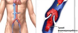
Thrombophlebitis of the saphenous veins of the mammary glands
Thrombophlebitis of the saphenous veins of the mammary glands is usually associated with thrombophlebitis of the superficial veins of the anterolateral region of the chest. Patients complain of painful tension on the hyperemic area of the skin.
Breast cysts
A breast cyst is a pathological cavity filled with liquid contents. If there is a breast cyst, a woman may feel aching pain, which is associated with an increase in the formation, which begins to compress the surrounding tissues.
Breast tumors
Pain appears in the presence of large or rapidly growing breast tumors, the addition of edema, inflammation, and decay. Benign tumors include adenoma, lipoma, hamartoma, etc., and malignant tumors include cancer and sarcoma.
Lactostasis
Lactostasis is the stagnation of milk in the mammary ducts of a woman’s glands during breastfeeding. A woman feels discomfort during and after breastfeeding. With lactostasis, pain, lumps in the mammary gland, a feeling of fullness and heaviness occur. Skin hyperemia develops and body temperature rises.
Developmental defects, breast anomalies
Among the malformations of the mammary glands, there are polymastia (additional mammary glands), polythelia (additional nipples), and mastoptosis (prolapse of the mammary glands).
If there are additional mammary glands or nipples, a woman may experience severe pain in the premenstrual period, as well as during pregnancy and lactation, if the glands cannot secrete milk. In drooping mammary glands, blood circulation is disrupted, swelling and lymphostasis form, which causes pain.
Symptoms of mastopathy
Most women consider moderate tenderness of the mammary glands before menstruation to be normal and do not consult a doctor with complaints. The mammologist is visited only by those whose breasts cause pain even when wearing a bra; they can only sleep on their backs, and if they have to travel in public transport, the slightest shock causes severe pain. A woman experiences pain when touching her breasts during sex. Only in this case do patients come to their senses and make an appointment with a mammologist.
In addition to pain, the woman feels an enlargement of the mammary glands, sometimes clear liquid is released from the nipple. The breast may feel heterogeneous to the touch, and sometimes lumps can be felt.
Diagnosis of the causes of pain in the mammary gland
- Consultation with a mammologist. After a thorough examination, the doctor can refer the patient to other specialists at our medical center - a gynecologist, endocrinologist, surgeon, oncologist, geneticist.
- Instrumental studies: ultrasound of the mammary glands;
- ductography (determining the patency of the ducts);
- digital and MR mammography.
- biopsy followed by histological examination;
Treatment of chest pain
It is extremely important to immediately go to a specialist when the first chest pain appears. If the symptom is provoked by any disease, such efficiency will prevent its intensive development.
It is equally important to find a truly competent specialist who works with proven and reliable diagnostic equipment. It is the accuracy of the latter’s testimony that largely determines how accurate the diagnosis will be made and, therefore, the optimal course of treatment will be selected.
A large staff of highly qualified therapists, gynecologists, oncologists, surgeons and ultrasound diagnostic specialists is assembled at JSC “Medicine” (clinic of Academician Roitberg). Conveniently located in the center of Moscow, the medical center building has many comfortable access roads, in particular, it is distinguished by the close location of the Tverskaya, Chekhovskaya, Novoslobodskaya, Belorusskaya and Mayakovskaya metro stations.
Reception of specialists is carried out by appointment. To get a consultation, just call 24/7 +7 (495) 775-73-60 or leave a request for an appointment in the return appointment form on the main page of the clinic’s website.
Treatment of breast pain
Treatment of painful sensations in the mammary gland is aimed at the underlying disease. The Clinical Hospital on Yauza provides treatment for identified mammary gland pathologies of any complexity:
- conservative drug treatment of breast diseases;
- surgical treatment, which includes sectoral resection of the mammary gland (organ-sparing surgery, without breast removal); radical mastectomy with removal of regional lymph nodes; plastic surgery (breast resizing, reconstruction, lift).
- chemotherapy - used in the presence of malignant neoplasms, breast cancer.
If you have any doubts about the health of the mammary glands, do not hesitate to consult a specialist mammologist. Even breast cancer detected at stage 1 is completely curable in 80% of cases and allows you to save the breast.
Causes of mastopathy
The main cause of mastopathy is a hormonal disorder, namely excessive production of estrogen, which promotes tissue proliferation, and a lack of progesterone, which regulates the proliferation process. The second reason may be an excessive amount of the hormone prolactin, which is responsible for milk production during breastfeeding.
Therefore, before prescribing treatment, you should establish the cause: take hormone tests.
Those women who have the following diseases are at risk:
- Ovarian tumors
- Myomas
- Endometriosis
- Inflammation of the pelvic organs
- Thyroid diseases
- Obesity
- Adrenal gland dysfunction
- Diabetes
- Diseases of the liver and gastrointestinal tract
Hormonal imbalance and, as a result, mastopathy can be caused by the following reasons:
- Chest injuries
- Tight, uncomfortable underwear
- Stress
- Irregular sex or lack thereof
- Abortion
- Refusal of breastfeeding (BF)
- GW less than 6 months and more than one and a half years
- Iodine deficiency
- Alcohol abuse
- Smoking
Pain in the mammary glands not associated with breast disease
Tietze syndrome. Tietze syndrome is a rare inflammation of the costal cartilage at the junction with the sternum. From 1 to 4 ribs are affected, the syndrome is masked as pain in the armpits or breast area. The cause of the disease is rib injuries, heavy physical activity, and exhausting training. The only way to make an accurate diagnosis is to undergo an X-ray or CT scan, which can help see these types of changes. A general and biochemical blood test, effective in diagnosing other pathologies, does not reveal pathologies in Tietze syndrome.
Wearing the wrong underwear. A woman's breasts change as they gain or lose weight. For every 5 kilograms gained, 100 grams of weight falls on the chest. If a woman has gained 10 or 20 kg (and after giving birth, it is quite possible that she has gained 25-30 kg), then instead of the usual bra size 80B, she needs to switch to 95D. In winter, the weight gains up to 5 kg, and a summer bra is no longer suitable. Wearing tight underwear leads to chest pain and discomfort.
With age, breasts also undergo changes. If glandular tissue predominates in youth, then with the onset of menopause it is replaced by adipose tissue, and the breasts stretch. In this case, a minimizer comes to the rescue. This is special underwear that evenly distributes the load on the shoulders and spine, giving the breasts a beautiful shape.
Diagnostics
A mammologist is involved in determining the cause of pain in the mammary glands; if necessary, the doctor involves other specialists for consultation. An integrated laboratory and instrumental approach allows for a detailed examination of not only the breasts, but also the endocrine system and the musculoskeletal system. The most informative studies are:
- Ultrasonography
. Breast ultrasound is indicated for young women, since the glandular parenchyma of the breast is denser and better visualized using sonography. Limited round formations or diffuse changes are detected, which are associated with the appearance of pain in the mammary gland. Identification of a cavity with thick walls and hypoechoic contents indicates the presence of an abscess. - Mammography
. An X-ray is used as a screening method in women over 40 years of age, and in young patients it is used to clarify the location and size of the tumor. Mammography allows you to study the contours and internal structure of the tumor; in inflammatory processes, widespread nonspecific structural changes predominate. - Radiography
. An X-ray of the spine is performed to assess the condition of the vertebrae and identify pathological displacements or deformities. If osteochondrosis is suspected, the method is supplemented with tomography (CT or MRI), which clearly visualizes the intercostal discs and cartilage tissue. Plain radiography of the chest cavity is required to search for tumor metastases. - Biopsy
. Severe pain in the mammary gland and detection of a neoplasm by instrumental methods serves as the basis for a fine-needle biopsy under ultrasound guidance. For greater information, a thick-needle trephine biopsy is performed. The resulting material is sent for histological and cytochemical examination. - Lab tests
. General and biochemical blood tests can reveal changes in the number of leukocytes and an increase in ESR. A specific diagnostic method is the determination of tumor markers and autoantibodies in the blood. To verify the bacterial cause of the disease, the discharge from the gland is cultured on selective nutrient media. - Additional methods
. If pain in the mammary gland can be caused by intercostal neuralgia, electroneurography is recommended. The study helps to study the excitability of nerve fibers and the speed of impulse transmission. Be sure to study the female hormonal profile on different days of the menstrual cycle.
Examination of the breast by a mammologist
Diseases of the respiratory system
Pathologies of the pleura and pleural cavity, as well as the lungs themselves, can provoke pain in the chest. A distinctive feature of these diseases is the presence of a connection between pain and breathing: it increases with inhalation and decreases with exhalation. This causes an involuntary decrease in breathing rate - it becomes frequent and superficial. Most often, chest pain in such situations is caused by pneumonia, pleurisy, pleural empyema and tumors of the bronchopulmonary localization and pleura.
Pneumonia
Pneumonia is inflammation of the lungs that occurs as a result of damage to the lung tissue by a variety of bacteria, viruses or fungi. Most often, it is a consequence of acute respiratory infections, including infection with a new strain of coronavirus, which is accompanied by a deterioration in the patient’s condition after a temporary improvement.
As a result, pneumonia has typical symptoms characteristic of most respiratory infections:
- rapid increase in body temperature to 38 °C and above;
- chills;
- headache;
- decreased appetite;
- cough is initially dry, and then with sputum, less often hemoptysis;
- dyspnea;
- superficial or deep pain in the chest, which tends to intensify with coughing and breathing;
- muscle weakness, joint discomfort;
- general weakness, depression.
Today, diagnosing pneumonia is not difficult and is usually detected during the initial examination of the patient.
Pleurisy
Pleurisy is inflammation of the pleural layers covering the surface of the lungs and chest wall. This is accompanied by the formation of fibrinous deposits or the accumulation of liquid exudate in the cavity formed by two layers of the pleura. This may be a consequence of an infectious lung infection, in particular tuberculosis, although it also occurs with bacterial, viral and fungal infections of other origins. Also, pleurisy can develop against the background of rheumatism, after myocardial infarction, as well as with gastrointestinal pathologies and in some other cases.
There are dry and exudative pleurisy, and the second form is often a consequence of untimely treatment of dry pleurisy. In such cases the following are observed:
- intermittent chest pain, aggravated by deep breathing, coughing, swallowing, and in some cases, by tilting the body in the opposite direction;
- pain radiating to the stomach, shoulder;
- severe, prolonged hiccups;
- lag of the affected half of the chest when breathing;
- dry cough;
- dyspnea;
- weakness, decreased performance;
- increase in body temperature to subfebrile values;
With pleurisy, there is often a friction noise between the layers of the pleura, which resembles the creaking of snow underfoot. Moreover, if you place your palm on your chest, you can feel their friction.
With exudative pleurisy, pain is present at the beginning of the development of the disease and at the end. But after a sufficient volume of exudate accumulates in the pleural cavity, they disappear. The remaining symptoms are generally similar to those of dry pleurisy.
Empyema of the pleura
Pleural empyema, in fact, is one of the variants of the course of pleurisy, but unlike other forms, it is characterized by the accumulation of pus in the pleural cavity. It can be a consequence of pneumonia (pneumonia), the formation of tumors, cysts, bronchiectasis and other disorders of the lungs, less often in the abdominal organs.
The pathology is characterized by:
- chest pain;
- dyspnea;
- fever;
- chills;
- decreased appetite;
- forced position of the body (lying on the sore side).
The development of pleural empyema requires hospitalization in the surgical department.
Tumors of the lungs and pleura
There are many types of benign and malignant neoplasms that can affect the lungs, bronchi and pleura. They are often accompanied by recurrent pleurisy with constantly increasing pain on the affected side. But direct chest pain caused by a neoplasm, especially a malignant one, occurs after it reaches a large size. This significantly worsens the prognosis, although today there are methods for treating even large tumors that were previously considered inoperable.
Diseases of the cardiovascular system
Pathologies of the heart and blood vessels still remain among the most common, which is largely due to the lifestyle of modern people. The most common causes of chest pain are:
- angina pectoris;
- myocardial infarction;
- pericarditis.
Angina pectoris
Angina pectoris is one of the clinical forms of coronary heart disease (CHD). It is a consequence of a decrease in the amount of oxygen entering the heart muscle (myocardium) due to impaired blood flow in the arteries (ischemia). This may be the result of the development of atherosclerosis of the coronary arteries, which leads to a decrease in their lumen, spasm of blood vessels or thrombosis and other disorders. The consequence in all cases is a deficiency of oxygen in the myocardium, which provokes the occurrence of characteristic symptoms:
- dull, pressing or pulling pain behind the sternum (sometimes it is localized in the region of the heart), tending to radiate to the left shoulder and the inner surface of the left arm, less often to the neck, right arm and stomach and passes when physical activity stops and the transition to a calm state, so attacks usually last less than 5 minutes;
- dyspnea;
- increased diastolic (lower) blood pressure.
A distinctive sign of angina pectoris is the occurrence of an attack during physical activity, transition from heat to cold, especially in strong winds. But the most pathognomonic symptom is the rapid elimination of pain after taking nitroglycerin.
Myocardial infarction
Myocardial infarction is a condition that requires emergency medical attention. It is a consequence of the progression of coronary artery disease, and in some cases the disease begins with it. This is a severe complication of myocardial ischemia, in which foci of necrosis, i.e., tissue death, form in it. This most often results from rupture of an atherosclerotic plaque. This triggers a whole chain of changes, the result of which is occlusion (blocking) of the coronary artery, which leads to the cessation of nutrition of the corresponding section of the myocardium and the development of ischemic necrosis.
Myocardial infarction is accompanied by severe, excruciating pain behind the sternum, which in nature can be either pressing, bursting or squeezing, or burning, stabbing, or felt like a blow to the heart. It is in many ways similar to an attack of angina, but is much stronger and more often accompanied by a fear of death. The pain is localized in the area behind the sternum and usually spreads to the left, less often it covers the entire surface of the chest and radiates to the stomach, reminiscent of appendicitis, exacerbation of gastric ulcer, etc.
Pain during myocardial infarction radiates to the left shoulder, forearm, hand, and more often it radiates to the little finger and ring finger. They can also radiate to the right hand or to the stomach area, under the shoulder blades, the left side of the neck, jaw, or ear.
The attack can last 15-30 minutes, but more often it drags on for several hours up to 24 hours. In addition to chest pain, this may be accompanied by:
- decreased blood pressure;
- nausea, vomiting;
- shortness of breath;
- severe weakness, combined with increased emotional arousal due to fear of death;
- dizziness;
- cold sweat;
- feeling of heartbeat;
- headache;
- pallor, marbling of the skin;
- tremor.
Shortly before the onset of pain, the following may be observed:
- varying degrees of chest discomfort or pain, as with angina pectoris, but occurring against the background of complete calm or with little physical activity;
- dyspnea;
- weakness;
- dizziness.
Myocardial infarction differs from an attack of angina pectoris in the severity of pain and the duration of its persistence. Also, during a heart attack, taking nitroglycerin leads to only a slight improvement or has no effect at all.
Pericarditis
Pericarditis is a disease accompanied by inflammation of the pericardial sac. Most often, its development is caused by concomitant disorders (viral, bacterial, fungal infections, in particular tuberculosis and borreliosis, as well as metastatic lesions, metabolic disorders), although it can also be a consequence of exposure to ionizing radiation, trauma, or occur in the absence of visible signs. causes (idiopathic pericarditis).
There are acute and chronic pericarditis. In the first case, the following are observed:
- pressing pain in the chest in the area of the heart of varying degrees of intensity, sometimes radiating to the left arm;
- increased pain when taking a deep breath, coughing and swallowing;
- reduction of pain when taking a sitting position, slightly leaning forward, and intensification in a supine position.
With chronic pericarditis, moderate chest pain and shortness of breath are observed. Often, patients previously noticed an increase in body temperature, weakness, muscle discomfort and other typical symptoms of infectious diseases.
With pericarditis, pain is relatively constant, and its severity increases gradually. Taking nitroglycerin does not eliminate the ailment, although NSAIDs (ibuprofen and others) help improve well-being.


