Rectum
The rectum is a tube with muscular walls consisting of longitudinal and transverse layers, it is fixed in the pelvis by ligaments and has a length of about 15 cm. The rectum then passes through the rectosigmoid junction into the sigmoid colon. The rectosigmoid junction has the appearance of a bend of 70-120 degrees. For 10-15 cm from the anus, the rectum has practically no painful endings and does not contact the small intestine located in the abdomen. Due to these features, when performing sigmoidoscopy, discomfort occurs only when it is necessary to examine the rectosigmoid section and the final segment of the sigmoid colon and is caused by passage through the rectosigmoid junction and contact through the wall of the sigmoid colon with the small intestine, on the surface of which there are many pain receptors, while diagnosis of the rectum itself is practically painless.
Causes of fistulas
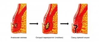
If you experience symptoms characteristic of a rectal fistula, in the vast majority of cases the cause for this was an inflammatory process in the anal gland (Fig. 2). In turn, inflammation of the gland is usually preceded by microtrauma of the anal canal. The cause of such injury is most often excessive irritation and/or trauma to the anal canal when too hard stool passes through the anal canal during constipation or, conversely, during diarrhea. Relatively rare causes of so-called specific fistulas of the anal canal and rectum are inflammatory bowel diseases (Crohn's disease, ulcerative colitis), tuberculosis, rectal tumors, and the consequences of radiation therapy of the pelvic organs. Inflammation of the gland is accompanied by the development of an abscess (ulcer) in the soft tissues of the perineum and is usually characterized by severe pain, which forces the patient to consult a doctor. Timely and correctly performed opening of the abscess leads to rapid relief of the condition, however, according to modern data, about half of all abscesses are accompanied by the subsequent formation of a fistula and require repeated intervention.
It is this repeated intervention that is aimed at radically getting rid of the fistula and requires not only an extremely thoughtful approach to the choice of treatment tactics, but also extensive surgical experience on the part of the surgeon.
Important! Opening an abscess is a relatively simple operation, but if performed incorrectly, it is at this stage that the muscular complex of the anal canal is most often damaged, which leads to disruption of the function of controlling intestinal contents!
If your visit to the doctor is postponed, the abscess may open on its own with the release of pus, after which your health usually improves and the pain in the perineal area decreases sharply. Symptoms may subside or disappear completely for a period of several weeks or months to many years. Despite the improvement in general well-being, spontaneous opening of an abscess is a relatively unfavorable scenario, because in this case, there is no complete evacuation of the contents of the purulent cavity, but only part of the purulent discharge occurs through the formed hole, which can soon close again. Proper treatment of an abscess consists of creating adequate drainage by creating a sufficient hole, evacuating the contents, rinsing the abscess cavity with antiseptic solutions and, if possible, removing the inflamed gland that is causing the inflammation.
Important! Sometimes, in the hands of an expert surgeon, with the relative simplicity of the fistula tract, radical excision of the fistula at the stage of surgical opening of the abscess is possible, but performing this intervention is extremely dangerous in the hands of an insufficiently experienced specialist and today is not recommended in most international recommendations.
When an abscess spontaneously opens, the likelihood of a return of the acute disease increases and the process becomes chronic with the formation of a fistula. A long course of the disease with periodic exacerbation leads to scarring of the surrounding tissues and, most importantly, the sphincter muscles responsible for the holding function. In addition, in conditions of constant inflammation, a relatively simple fistula can give rise to additional passages and leaks, which significantly complicates subsequent treatment.
Timely consultation with a doctor at an early stage of the disease can reduce the likelihood of developing a fistula and such dangerous complications of the inflammatory process as pelvic phlegmon. Therefore, even minor symptoms that do not significantly affect the quality of life require attention and evaluation by a qualified specialist.
Anal canal
The anal canal is the terminal part of the rectum, it ends with the anus and has a pronounced muscular wall - the anal canal sphincter complex. This complex consists of external and internal sphincters, which are responsible for voluntary and involuntary retention of feces and gases. In the anal canal, the rectal mucosa transitions into the epithelium (anodermis) of the anal canal, which then passes into the skin of the perianal area. In the skin part of the anal canal there is a very large number of pain receptors, and on the mucous membrane they are practically absent. When an anal canal fissure develops, it is located on the skin part, its bottom is the subcutaneous/superficial portion of the sphincter (constituting no more than 1/10 of its total volume), and with mechanical irritation (bowel emptying), a pronounced spasm of this muscle develops, and intense pain syndrome. That is why performing a drug blockade or dosed sphincterotomy in the case of a fissure with pain that is not amenable to conservative treatment leads to its rapid healing.
Symptoms of acute paraproctitis depend on the localization of the process, the type of pathogenic microorganisms and the reactivity of the body. The disease begins acutely after a short prodromal period with malaise, weakness, and headache. Fever, headache, and increasing pain in the perineum and pelvis appear. If the inflammatory process in the perirectal tissue is not limited and proceeds like phlegmon, septic signs occur. As the abscess forms, the pain increases and becomes pulsating. This period ranges from 2 to 10 days. Subsequently, the abscess breaks into the rectum or onto the skin of the perineum. In 70% of cases, a breakthrough of the abscess is manifested by a short-term improvement in the condition. In 30-70% of cases, acute paraproctitis becomes chronic. If an acute inflammatory process occurs against the background of fistulas, then this form is called chronic recurrent paraproctitis. After opening the abscess, the internal opening remains open. The hole in the skin does not close, and bloody or purulent discharge periodically appears from it. Temporary closure of the internal opening leads to remission. The period of temporary well-being can last several months, or even years. Subcutaneous paraproctitis. This is the most common form, accounting for 50% of all types of paraproctitis. Patients complain of rapidly increasing pain in the anus, perineum, pulsating in nature, intensifying with changes in body position, coughing, and defecation. There is stool retention. When the abscess is localized in front of the rectum, dysuric phenomena may occur. Body temperature rises to 38-39°C with chills.
On examination: the skin of the perineum on the affected side is hyperemic. The radial folding at the anus is smoothed out. A protrusion of the skin appears, which takes on a spherical shape. When the abscess is located in the anus, the latter becomes deformed, becomes slit-like, and sometimes gapes. In such cases, gas incontinence, liquid feces, and mucus leakage occur. Palpation is sharply painful. In 50% of cases fluctuation is determined. A digital examination of the rectum reveals a painful infiltrate and a smoothed anal canal. Instrumental research is extremely painful and even impossible. Acute submucosal paraproctitis is a mild form of paraproctitis. Occurs in 2-6% of cases. Patients complain of mild pain in the rectum, which intensifies during defecation. Within a week, pus, as a rule, breaks into the lumen of the rectum and recovery occurs. Upon examination, submucosal paraproctitis manifests itself when the process extends below the pectineal line (swelling of the corresponding semicircle of the anus). A digital examination reveals a painful, round, tightly elastic formation under the mucous membrane above the pectineal line. Ishiorectal paraproctitis. Occurs in 35-40% of cases. Initially, patients complain of a deterioration in their general condition, fever, and poor sleep. Subsequently, vague heaviness and dull pain in the rectum appear. By the end of the 1st week, the patient's condition worsens. The temperature rises to 39-40°C. The pain becomes acute, throbbing, intensifies with defecation or sudden movements. When the abscess is localized in the area of the prostate or urethra, dysuric disorders occur.
On the 5-7th day of illness, swelling, swelling, and slight hyperemia of the skin of the perineum appear,
How to behave after surgery?
After the operation, you will remain in the hospital for 1 to 5 days, depending on the complexity of the operation. You will be prescribed, if necessary, antibacterial therapy and dressings with wound-healing ointments will be performed. After surgery, you may need to hold stool to allow the wound to heal.
Your doctor will teach you how to care for the wound, and it is extremely important to follow these recommendations strictly. One of the important recommendations is to irrigate the wound with a stream of water 3-4 times a day for the purpose of mechanical cleaning and periodic medical monitoring of proper healing in the direction “from the bottom of the wound.” After training, as a rule, you will be able to perform dressings yourself at home.
Which doctor should you contact if symptoms occur?
If you or your loved ones have the described complaints, you should contact a coloproctologist as soon as possible, who, after consultation and examination, will establish a preliminary diagnosis and determine further tactics and urgency of treatment, or, if in doubt about the diagnosis, a list of necessary additional studies.
If surgery is required, you will be offered hospitalization in a hospital, where, after assessing the results of the studies, which, as a rule, do not take much time and are easy to perform, the operation will be performed.
Anatomy of the rectum, obturator apparatus and pararectal tissue spaces
Rectum
The length of the rectum is on average 15 cm. Traditionally, its two main sections are distinguished: pelvic and perineal, the first lies above the pelvic diaphragm, and the second below. A.N. Ryzhikh (1960) distinguishes three sections of the rectum: perineal, ampullary and rectosigmoid [16]. According to M.G. Prives (1974), the rectum is more conveniently divided into five sections: perineal, lower ampullary, mid-ampullary, upper ampullary and supramullary (rectosigmoid), the approximate length of each of them is 3–4 cm [14]. Foreign sources that describe the surgical anatomy of the rectum divide it into three sections: the upper third, the middle third and the lower third, the length of each section is approximately 5 cm [18,19].
The anatomical boundaries of the rectum also remain controversial. Some authors consider the third sacral vertebra to be the proximal border of the rectum [13,16], others – the promontorium [14]. There is also no unanimity on the issue of the identity of the rectosigmoid colon. In Russian literature, it traditionally refers to the rectum and is called the supramullary rectum [1, 13, 16]. In foreign sources, the rectosigmoid section of the colon is called the rectosigmoid junction and is interpreted as an independent section of the digestive tract about 3 cm long, the signs of which are the gradual loss of the mesentery and haustration and the fusion of the free and omental tena into a single anterior [17]. In the International Classification of Diseases ICD-10, the rectosigmoid section of the colon is also called the rectosigmoid junction and is considered as an independent anatomical unit.
Rice. 1. Anatomy of the rectum: 1 – serous membrane (peritoneum) of the rectosigmoid colon; 2 – ampulla of the rectum; 3 – anal canal; 4 – internal sphincter of the anus; 5 – external anal sphincter; 6 – anus; 7 – anal ridge; 8 – anal columns of Morgagni; 9 – anal sinus; 10 – muscle that lifts the ani; 11 – transverse fold of the rectum; 12 – mucous membrane; 13 – muscular layer
The rectum is characterized by the absence of shadows, fatty pendants, haustrations and mesentery. The upper third of the rectum is covered with peritoneum anteriorly and laterally, the middle third is only anteriorly, and the lower third is located extraperitoneally - under the pelvic peritoneum (Fig. 1).
In the distal part of the anal canal, Hilton's white line is identified - the line of attachment of the distal fibers of the rectal levators. In this area, a groove can be detected by palpation [16], distal to which the anal mucosa gradually passes into the skin [15]. The skin epithelium in this area is thinned, smooth, pale, devoid of hair follicles and glands. Even further distal, hair, sebaceous and sweat glands appear, which already characterizes the skin as perianal. The anal mucosa has longitudinal depressions - Morgagni crypts, into which the mouths of the anal glands open, numbering from 4 to 12. The crypts are separated by longitudinal folds or Morgagni columns, their number ranges from 8 to 14. At the base of Morgagni columns there are anal papillae in the form of small elevations . Normally, they do not exceed 2–3 mm, but in some cases they can hypertrophy up to several centimeters in length. In the same area, a dentate line is also defined, representing the border of the squamous epithelium of the anus and the transitional epithelium of the cloacogenic zone of Morgagni columns. The last zone has a length of 0.5 to 1 cm; above it is the columnar epithelium of the rectal ampulla.
The internal anal sphincter is a thickened distal 3–4 cm of the circular smooth muscle layer of the rectum. Being in continuous tonic tension, it is a barrier to the involuntary evacuation of feces and gases. The external anal sphincter is a striated muscle that encircles the anus in a cone-shaped manner, tapering downwards. The sphincter consists of three portions: subcutaneous, superficial and deep. All of them are separated from each other, as well as from the internal anal sphincter, by thin fibrous septa, through which the fibers of the rudimentary connecting longitudinal muscle, woven into the perianal skin, pass from top to bottom. The deep portion of the sphincter is anatomically (but not embryologically) connected with the puborectal muscle and the levator muscle.
Rice. 2. Cavities around the rectum filled with fatty tissue (Littmann I.): 1 – spatium ischiorectale, 2 – spatium pelvirectale, 3 – spatium perianale
The levator ani muscle is the pelvic diaphragm, or the main element of the pelvic floor. It consists of a pair of broad, symmetrical fibers that are divided into three main components: the iliococcygeus muscle, the pubococcygeus muscle, and the puborectal muscle. It is also possible that there is a variable fourth component - the rudimentary ischiococcygeus muscle, represented by several muscle fibers on the surface of the lig. sacrospinale.
Pararectal spaces
The following clinically significant peri-rectal spaces are distinguished: ischiorectal, perianal, intersphincteric, submucosal, superficial postanal, deep postanal, supralevator and retrorectal. The first two spaces - ischiorectal and perianal - are separated from each other by a thin horizontal fascia. The upper two-thirds of the ischiorectal fossa is made up of the ischiorectal space, located on both sides of the rectum, having a pyramidal shape and limited medially by the rectum, laterally by the side wall of the pelvis, and superiorly by the levator (Fig. 2). The pudendal nerve and the internal pudendal artery and vein pass superolaterally. The inferior rectal artery and vein are also found in the tissue of the ischiorectal fossa. At the base of the pyramid described above is the perianal space, which surrounds the lower part of the anal canal and extends laterally to the subcutaneous fatty tissue of the buttocks, and medially to the intersphincteric space. It is also limited by the subcutaneous portion of the external sphincter, the lower part of the internal sphincter and the fibers of the longitudinal muscle. In the perianal space there is an external hemorrhoidal plexus, communicating with the internal one in the area of the dentate line. This area is a typical site for the formation of perianal hematomas, fistulas and abscesses.
Treatment before and after surgery
Chemotherapy and radiation therapy are indicated for patients with stage 2 or higher tumors.
If before the operation metastases were detected in several lymph nodes, and the tumor has grown into the muscle layer, then at the stage of preparation for the operation a short course of radiation therapy is carried out for 5 days. This allows you to destroy early metastases and reduce the size of the formation itself.
Treatment of rectal cancer after surgery is carried out after obtaining pathomorphological data on the removed tissues. The issue of radiation or its combination with chemotherapy is being decided. Radiation therapy after surgery destroys the remaining cells in the area of the primary tumor and prevents its recurrence. In inoperable patients, it alleviates the condition.
Sensitivity to chemotherapy is detected in 30% of patients. It is prescribed for therapeutic purposes to destroy metastases.
Chemotherapy is also carried out adjuvantly - to prevent the spread of carcinoma if damage to several lymph nodes is detected. This method of therapy improves the quality and life expectancy of patients with metastases. Platinum preparations, 5-fluorouracil, leucovarin, and calcium folinate are used. Medicines are administered intravenously in courses of several days. Chemotherapy is also used in combination with radiation before surgery for locally advanced cancer. This combined treatment is carried out for 1-1.5 months, and after the end of irradiation, surgery is performed 6 months later.
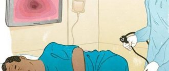
What is this procedure?
This is a diagnostic examination of the rectum, the essence of which is a visual examination of the walls of the rectum through an endoscope. This device is equipped with a high-quality camera that transmits the image to the monitor. Thanks to this, the doctor can assess the condition of the organ from all sides, study its structure and correct functioning. This procedure is prescribed not only for diagnostic purposes, but also for treatment purposes. The structure of this device makes it possible to simultaneously carry out minor surgical interventions (removal of polyps, stenting, stopping bleeding, removal of foreign bodies, etc.).
Introduction
Paraproctitis - acute or chronic inflammation of the peri-rectal tissue - is a very common disease. In the structure of proctological diseases, it ranks fourth after hemorrhoids, anal fissures, and colitis. Among people with diseases of the rectum, patients with acute paraproctitis account for an average of 15.1%. Paraproctitis is more common in males, mainly in working age (from 20 to 50 years).
Mention of this suffering dates back to the era of Hippocrates, but the treatment of paraproctitis has its own characteristics and remains a difficult task even today.
Patients with acute paraproctitis often end up in hospitals on duty with general surgeons, so their competence in diagnosing and treating this pathology is extremely important.
In this publication, we have tried to present modern methods of diagnosis and treatment of paraproctitis in an accessible manner.
Treatment of complex fistulas should be divided into 2 stages
At the first stage, a drainage ligature is installed into the fistula tract - a thin, almost invisible suture material (Fig. 7). The main goals of this stage of treatment are: the formation of a direct fistula tract around the ligature, drainage of possible leaks, reduction of the inflammatory process, prevention of self-closure of the external opening for constant drainage of inflammatory and/or purulent discharge.
Figure 7. Drainage ligature
At the second stage, after the acute inflammation subsides (4-6 weeks), the main surgical treatment is performed - excision of the fistula.
Important! Two-stage surgical treatment provides conditions under which radical surgery becomes possible with minimal complications.
Currently, in the treatment of rectal fistulas, various biological materials based on connective tissue (Permacol), fibrin glue, and collagen paste are widely used, which are introduced into the fistula tract, tamponing it. These methods have a lesser effect on the rectal closure apparatus; their use does not lead to the formation of a massive wound defect. However, the rate of disease recurrence is significantly higher than radical surgery.
Promising modern methods for treating fistulas are endoscopic (VAAFT) and laser (FiLaC) technologies for removal of affected tissue. These technologies practically eliminate the possibility of damage to the sphincter apparatus, however, today, the effectiveness in leading clinics in the world does not exceed 50-60%.
