Consultation with a proctologist – 1,750 rubles.
- What is paraproctitis (rectal fistula)?
- Symptoms of paraproctitis
- Types and stages of the disease
- Diagnosis of rectal fistulas
- Treatment of paraproctitis
- Surgery
- Main risks
- Prevention
- Popular questions
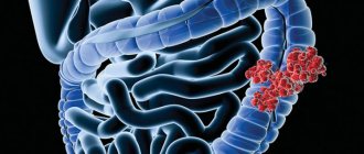
Chronic paraproctitis (rectal fistula)
– a chronic inflammatory process in the anal crypt, intersphincteric space and pararectal tissue with the formation of a fistulous tract. The affected crypt is the internal opening of the fistula.
According to statistics, most patients with rectal fistulas associate the occurrence of the disease with acute paraproctitis. About a third of patients with acute paraproctitis consult a doctor after spontaneous opening of an abscess, after which in most cases a rectal fistula is formed. Approximately half of the patients with acute paraproctitis undergo radical surgery at the time of treatment, the other half only undergo opening and drainage of the abscess without eliminating the entrance gate of the infection, which can subsequently lead to the formation of a rectal fistula. In this case, constant infection occurs from the intestinal lumen, and a fistula tract is formed from scar tissue. The external opening of the fistula is most often localized on the skin of the perineum, its diameter is often no more than 1 mm. If the fistula tract is insufficiently emptied, purulent cavities may form along its length.
The prevalence of chronic paraproctitis reaches 23 cases per 100,000 population, the proportion among rectal diseases reaches 25-30%. Males are more susceptible to this disease than women; the disease more often manifests itself in young and middle age Source: Khomochkin V.V. Operation of ligation of the fistula tract in the intersphincteric layer in the treatment of chronic paraproctitis / V.V. Khomochkin [et al.] // News of surgery. - 2021. - T. 26. - No. 5. - P. 616-623. .
Symptoms of paraproctitis
Symptoms directly depend on where the fistula is located and the state of the patient’s immunity.
Usually, after an illness, a person experiences pain in the anus and a hole appears from which pus comes out. This causes itching, irritation, and bad odor. Sometimes ichor is released along with pus. This occurs when blood vessels are damaged. If the fistula has no outlet, then patients complain only of pain and/or discharge from the rectum or vagina.
An incomplete internal fistula provokes a sensation of the presence of a foreign body in the anal area. If the infiltrate does not leave the fistula cavity enough, the following symptoms appear:
- discomfort and pain in the anal area;
- retention of urination and defecation;
- discharge of mucus, infiltrate, pus from the rectum;
- increased body temperature, chills;
- redness and irritation of the skin around the anus and part of the buttocks.
Chronic paraproctitis during the period of exacerbation gives the following symptoms:
- fast fatiguability;
- sleep disturbance;
- nervous exhaustion;
- headache;
- regular increase in body temperature;
- intimate disorders;
- gas incontinence.
Physiological changes may occur:
- anal deformation;
- disruption of the anal sphincter;
- scars on the muscle tissue of the anal sphincter.
During periods of remission of chronic paraproctitis, the general condition is normal; with proper hygiene, the quality of life is almost not reduced. But with a long course of the pathology and frequent exacerbations, the following may occur:
- sleep disorders;
- powerlessness, weakness;
- headache;
- periodic changes in body temperature;
- nervousness;
- poor work ability;
- deterioration of potency.
Anesthesia
For surgical interventions in the perineal area, regional anesthesia is currently used. It includes subarachnoid (spinal) anesthesia at the lumbosacral level, which allows you to block the transmission of sensory and painful nerve impulses to the lower part of the body, including the anorectal area. In this case, the motor function of the lower extremities is not lost. Subarachnoid anesthesia is a fairly safe method and has a low risk of complications from the heart, respiratory system and nervous system. During the operation, for maximum comfort, the patient is conscious or in a state of medicated sleep (sedation). General anesthesia in anorectal surgery is used in individual cases at the discretion of the anesthesiologist-resuscitator.
Types and stages of the disease
Rectal fistulas can be complete, incomplete, or internal.
- Complete ones
have two openings - located inside and opening into the rectum, as well as external, usually located next to the anus. - An incomplete fistula
has only an internal opening that opens into the rectum. There is an opinion that this is a transitional phase, because sooner or later the external opening will open and the fistula will become complete. - An internal fistula
has two internal openings that extend into the lumen of the rectum.
According to their location relative to the anal sphincter, fistulas are divided into:
- Intrasphincteric
, or marginal. The most common ones usually have a straight course without scars and an external opening near the anus. - Transsphincteric
- the course of such a fistula goes through the anal sphincter at different depths. The higher the course, the more it branches and the more often purulent streaks form, and scar tissue forms around the fistula itself. Scars can deform the anal sphincter and interfere with its function. - Extrasphincteric
- differ in the location of the internal opening - on the surface of the intestinal crypt. The stroke itself passes high and goes around without affecting the outer pulp. The incidence of such fistulas is 15-20% of the total incidence.
Extrasphincteric fistulas are usually tortuous and have a long course. With them, purulent leaks form and scars form around the fistula canals. With repeated exacerbations, new external openings appear. There is a classification that distinguishes four degrees of complexity of fistulas of this type:
- – there are no scars around the internal opening, the course is straight, infiltrates and purulent streaks do not form in the perirectal tissue.
- – scars appear around the internal opening, there are still no ulcers or infiltrates.
- – the entrance to the fistula canal is narrowed, there are no scars, there are ulcers and inflammatory infiltrates in the perirectal tissue.
- – the inlet is enlarged, surrounded by multiple scars, and there are ulcers and infiltrates in the tissue.
Methods for diagnosing rectal fistulas
In the vast majority of cases, the diagnosis is not difficult to make. The proctologist is based on the patient’s complaints, examination and palpation data. Usually, during a digital examination, the doctor discovers holes in the anal area, from which pus comes out when pressed.
When diagnosing and subsequent treatment of rectal fistulas, the determination of the fistula tract is of fundamental importance. For this purpose, digital and mirror examination of the rectum, probing and examination of the fistulous tract with a dye solution, anoscopy or sigmoidoscopy, fistulography, ultrasound, CT, MRI are used Source: Seidova K.R. Etiopathogenesis and diagnosis of pararectal fistulas. Literature review / K.R. Seidova // Bulletin of surgery of Kazakhstan. - 2011. - No. 4. - P. 23-27. .
One of the main methods for studying pararectal fistulas is probing. With the help of probing, along with the internal opening of the fistula, it is possible to determine the depth of the fistula tract, its connection with the intestinal lumen, the relationship of the fistula to the anal sphincter, and by the movement of the tip of the probe in the canal, bay-shaped expansions and additional branches.
Examination of the rectum using a speculum and proctoscopy allows one to distinguish between altered crypts and the internal opening of the fistula. Injecting a dye into the external opening of the fistula allows you to more clearly distinguish the internal opening. For this purpose, a 1% aqueous solution of methylene blue, iodine, ink, manganese and other solutions are used.
Restoromanoscopy is an endoscopic technique that involves inserting a special tube equipped with optics into the anus. The doctor can assess the condition of the mucosa and do a biopsy to rule out a tumor if one is suspected.
Fistulography is a radiopaque technique. A contrast agent is injected into the anus, and then x-rays are taken. X-ray examination reveals the presence of pockets, cavities, extensive bays, additional branches and curved fistula tracts.
Transrectal ultrasound allows us to trace the fistula tract in the pararectal tissue, the presence of additional cavities along the course, the relationship of the fistula to the anal sphincter, determination of the internal opening of the fistula, differentiation from pararectal neoplasms. Ultrasound examination is also informative in determining anal sphincter insufficiency after surgery in patients with perirectal fistulas.
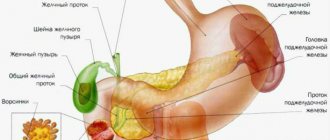
Women must receive a referral to a gynecologist for examination to rule out a vaginal fistula.
Complications
In modern conditions, subject to all standards of surgical technique, correct diagnosis and postoperative management, the risk of complications is minimal. But it cannot be said unequivocally that complications do not occur at all, therefore in each case one should treat this disease very carefully and carefully. The most common and most severe of all complications is fecal and gas incontinence. It develops as a result of damage by a pathological process or damage to the sphincter apparatus during surgery. Relapses (repeated cases of the disease), complicated forms of fistulas and the presence of acute inflammation pose a risk of developing incontinence. Another complication of rectal fistulas is bleeding. It can be immediately after surgery or a few days later. Usually it can be stopped in a dressing room by simply packing the wound. Otherwise, repeat surgery may be required.
Such a complication as failure or divergence of sutures on the mucomuscular flap occurs mainly in recurrent or complicated forms of rectal fistula. Due to the significant severity of scar tissue changes around the fistula tract, the tissues take longer to heal and are more susceptible to repeated inflammatory processes.
Treatment methods for paraproctitis
Complete cure is only possible through surgery. Physical therapy, antibiotics, and ointments are used in preparation for or after surgery. Drug therapy can also relieve symptoms and speed recovery. Main recommended pharmacological groups:
- painkillers;
- systemic antibiotics;
- healing ointments.
A course of physiotherapy may include ultraviolet irradiation and electrophoresis.
SM-Clinic specialists diagnose and treat complex pathologies. To make a diagnosis, the doctor collects the patient’s complaints, medical history and objective examination. To clarify the diagnosis and carry out differential diagnosis, additional instrumental studies are performed, such as anoscopy and sigmoidoscopy. These studies are carried out after cleansing the intestines by performing cleansing enemas and taking laxatives.
Surgical treatment of pararectal fistulas
The only indication for surgery is the presence of a fistula.
Contraindications:
- general serious condition of the patient;
- liver and kidney failure;
- chronic diseases in the stage of decompensation;
- blood clotting disorder;
- acute infections in the body.
The operation is usually carried out as planned. In case of exacerbation, the abscess is opened, and the fistula itself is removed after 1-2 weeks.
Preparation for surgery includes standard preoperative examination: blood tests, urine tests, ECG, fluorography. Additionally, rectal examinations and/or ultrasound may be prescribed.
There are several types of operations. A specific type is selected depending on the complexity of the pathology:
- ligature technique;
- excision of the fistula along its entire length with or without subsequent suturing;
- plastic surgery of the internal opening of the fistula;
- laser cauterization of the fistula tract;
- filling the fistula tract.
The surgical intervention is performed in a hospital; from 3 to 10 days after it it is necessary to remain under the supervision of doctors. The effectiveness of the operation reaches 90%.
Postoperative period
In the first days in the hospital, a gas tube is installed, antibiotics are prescribed, and dressings are performed. The pain syndrome is relieved with analgesics. It is important that there are no stools in the first 2-3 days, so a liquid diet is prescribed.
From the second or third day you can eat pureed, easily digestible food, you need to drink a lot. It is necessary to prevent constipation, which can cause the seams to come apart. Sitz baths with antiseptic solutions are prescribed, and, if necessary, laxatives and analgesic ointments.
After discharge, the patient should listen sensitively to his body and, if the following symptoms occur, immediately consult a doctor:
- excessive gas formation;
- fecal incontinence;
- a sharp increase in body temperature;
- constant abdominal pain;
- discharge of blood and/or pus from the anus;
- pain when urinating or defecating.
Basic recommendations for the rehabilitation period:
- eat small portions 6 times a day;
- drink at least 2 liters of water per day;
- do not consume foods that irritate the intestines - carbonated drinks, alcohol, chocolate, hot spices, fatty foods, foods with flavoring additives, etc.;
- give preference to vegetables and fruits as sources of fiber, eat cereals, grain bread and dairy products.
Why do you choose us?
Part of the Perm State Medical University named after. I.M. Sechenova Clinic of Coloproctology and Minimally Invasive Surgery is an example of a new generation of medicine, harmoniously combining the deepest fundamental knowledge, honed skills, a modern multidisciplinary approach and attentive attitude to any category of patients.
KKMH is a guarantee that the surgical interventions performed within our walls correspond to the most current ideas about colorectal surgery. This includes our own department of anesthesiology and intensive care, whose employees ensure a smooth course of the operation and the early postoperative period and treat seriously ill patients. This is an “open intensive care unit”, where you can not only find out the most complete information about the condition of a loved one, but also be with him during a difficult time for him.
The doors of our Clinic are open to patients who were denied treatment in other hospitals due to the complexity of the surgical intervention or the neglect of the process. Elderly patients, patients with a “bouquet” of concomitant diseases (so-called comorbid) are an area of our special interest. The presence in the hospital of such highly professional specialists as a cardiologist, pulmonologist, urologist, etc. allows us to treat patients of any age and with any concomitant diseases.
KKMH is a dynamically developing team of specialists who sincerely and deeply love their work, who are continuously learning and teaching others, who are interested in keeping you healthy.
The main risks associated with paraproctitis
Deep fistulas of the extra- and transsphincteric types often recur.
Long-term progressive fistulas, which are complicated by the process of scarring of the walls of the rectum and purulent leaks, can lead to secondary functional changes.
Periods of new exacerbations are dangerous due to the appearance of fresh foci of inflammation and the involvement of an increasingly larger array of the anal sphincter in the inflammatory process. All of the above inevitably affects the general condition of the patient. Often there are complaints of weakened immunity, headaches, sleep problems, decreased performance, and irritability.
In addition to the above complications, a long-existing fistula can lead to pectenosis, namely a cicatricial change in the wall of the anal canal with decreased elasticity and cicatricial stricture. With a long-term illness, in some cases, malignancy of the fistula tract is possible.
Causes
If the intestinal fistula is congenital, then it appears due to abnormalities in the development of internal organs. This may be non-closure of the bile duct, anomalies of the intestinal-umbilical duct. Pathology can also appear due to mechanical damage to the intestinal walls due to injury or surgery. Moreover, it is the operation that causes intestinal fistulas in half of the cases.
Traumatic damage to the intestine can occur with shrapnel or knife wounds, or blows to the abdomen. But this rarely happens in peacetime. But complications after surgery are quite common. This may be intestinal obstruction, incorrectly placed sutures, the appearance of abscesses, or prolonged unjustified drainage. Sometimes the pathology is caused by medical errors, for example, due to improper removal of the appendix, opening of abscesses or intubation of the small intestine. It can also be rough probing or leaving gauze swabs in the abdominal cavity.
Intestinal fistulas can also appear without mechanical damage due to necrosis of the intestinal wall. The reason for this may be various factors:
- disturbance of blood supply;
- long-term inflammatory process;
- Crohn's disease;
- acute appendicitis;
- the appearance of diverticula in the intestine;
- presence of foreign bodies;
- intestinal obstruction;
- peritonitis;
- strangulated hernia;
- intestinal tuberculosis;
- actinomycosis;
- cancerous tumors.