Right-sided oorphitis or inflammation of the right ovary in women has symptoms similar to appendicitis, peritonitis, and acute endometriosis. Therefore, sometimes diagnosing the disease can be difficult. The development of inflammatory processes in the right ovary is associated with the anatomical feature of the organ. Delayed diagnosis and treatment can cause severe complications, including complete loss of female reproductive function. At the Healthy Family multidisciplinary clinic, every woman in need of professional medical care can undergo the comprehensive diagnostics necessary to make an accurate diagnosis. If necessary, our specialists, taking into account the course of right-sided oorphitis, will prescribe effective treatment that will help you quickly recover and prevent the development of negative consequences.
Causes of inflammation of the ovary on the right side
Inflammation of the ovary on the right develops due to the penetration of pathogenic microflora from organs located nearby, such as:
- appendix;
- intestines.
When the appendix or rectum becomes inflamed, the infection spreads to adjacent tissues, affecting the ovaries. The right ovary is more susceptible to diseases than the left due to the anatomical features of the organ. The fact is that the gland located on the right is slightly larger in size than the left one. Also, the right ovary is more actively supplied with blood flow, and in the presence of a focus of infection, pathogenic microorganisms most often spread through the hematogenous route throughout all internal organs, affecting healthy tissue.
The causative agents of oorphitis can be:
- Opportunistic microflora that constantly lives in the body and is activated when the immune system is dysfunctional: staphylococci, streptococci, fungi.
- Infections that are sexually transmitted: gonococci, mycoplasmas, trichomonas, chlamydia.
The danger of right-sided inflammation is that after the infection penetrates the right ovary, it quickly spreads and spreads to the left, causing bilateral oorphitis, which often leads to disruption of the functioning of the female reproductive glands and the development of infertility.
Factors that can serve as a trigger for the occurrence of inflammatory processes in female paired glands:
- recurrent inflammatory diseases of organs located in the abdominal cavity;
- abortion, surgical interventions that injure the tissues of the reproductive system;
- systematic hypothermia, which reduces the body’s protective functions;
- promiscuous unprotected sexual intercourse;
- uncontrolled use of hormone-containing drugs;
- Exceeding the period of use of the intrauterine device.
Right-sided orphitis can occur as a result of the progression of distant infections that spread throughout the body along with lymph and blood. In this case, the causes of inflammation of the right ovary:
- advanced dental caries;
- chronic tonsillitis;
- appendicitis;
- cystitis, pyelonephritis.
External genitalia
1 – pubis; 2 – foreskin of the clitoris; 3 – head of the clitoris; 4 - labia minora; 5 - external opening of the urethra; 6 - hymen (is the border between the external and internal genital organs); 7 - Bartholin gland; 8 - anus; 9 — entrance to the vagina; 10 - labia majora.
Pubis
The pubis is an elevation located in front and slightly above the pubic joint, covered with hair, the upper limit of which grows horizontally (in men, hair growth extends upward along the midline).
Clitoris
The clitoris is a small (up to 1-1.5 cm), but very sensitive and important organ, consisting mainly of the corpus cavernosum. The male penis has a similar structure. The cavernous body has voids filled with circulating blood. During sexual arousal, these voids are intensely filled with blood, and the clitoris becomes enlarged and thickened—an erection. The corpus cavernosum is not capable of contracting like blood vessels, so traumatic injury to the clitoris is dangerous due to excessive bleeding.
Labia minora
The labia minora (LGB) are two folds of skin between the labia majora and the opening of the vagina. In front, connecting, they form the foreskin of the clitoris. Normally, the labia minora protrude slightly beyond the boundaries of the major lips, their color varies from pale pink to dark brown in the posterior regions. MPGs have a large number of vessels and nerve endings and are an ergenic zone; during sexual arousal, they increase in size due to blood flow.
PGMs are variable in shape and size, and are often asymmetrical. If the shape and size of the labia minora cause physical or mental discomfort, surgical correction of their size and the shape of the labia minora are performed.
sex slit
The genital fissure is the space between the labia majora and labia minora.
Labia majora
The labia majora (LGB) are two pronounced longitudinal folds of skin located on the sides of the genital slit. Anteriorly, the BPG converge into the anterior commissure, located above the clitoris. Behind, narrowing and converging one on the other, the BPG pass into the posterior commissure. The skin of the outer surface of the GPG has hair and contains sweat and sebaceous glands. The thickness of the labia majora contains blood vessels, nerves and Bartholin glands. On the inside they are covered with thin pink skin similar to a mucous membrane.
There are two openings under the labia majora and minora. One of them, with a diameter of 3 - 4 mm, located just below the clitoris, is called the external opening of the urethra (urethra), through which urine is discharged from the bladder. Directly below it there is a second hole with a diameter of 2 - 3 cm - this is the entrance to the vagina, which covers (or once covered) the hymen.
External opening of the urethra
The external opening of the urethra has a round, semi-lunar or star-shaped shape, it is located 2-3 cm below the clitoris. The urethra is 3-4 cm long, its lumen stretches to 1 cm or more. Along its entire length it is connected to the anterior wall of the vagina. On both sides of the external opening of the urethra are the excretory ducts of the paraurethral glands. These formations produce a secretion that moisturizes the mucous membrane of the external opening of the urethra.
Bartholin glands
Bartholin's glands (glands of the vestibule of the vagina) are paired, oblong-rounded formations, the size of a bean. They are located on the border of the posterior and middle third of the labia majora and produce a whitish secretion with a specific odor. The secret moisturizes the mucous membrane and has antibacterial properties.
Vaginal vestibule
The vestibule of the vagina is an anatomical formation. The “bottom” of the vaginal vestibule is the hymen or its remnants. In front, the vestibule is limited by the clitoris, behind - by the posterior commissure, on the sides - by the labia minora.
Hymen
The hymen (hymen) is the thinnest ring-shaped or semi-lunar-shaped membrane, 0.5 - 2 mm thick. With the onset of sexual activity, the hymen is torn. The hymen is the boundary between the external and internal genitalia.
Crotch
The perineum in the anatomical sense is the area between the pubis and the top of the coccyx, on the sides limited by the ischial tuberosities of the pelvic bones, in fact it is the exit from the small pelvis. In a clinical (obstetric) sense, the perineum is considered the area between the posterior commissure of the labia majora and the anus (Fig. 1). On the skin of the perineum there is a pigment line running from the posterior commissure to the anus - the perineal suture. The distance from the posterior commissure to the anus is called the perineal height and is 3-4 cm.
Rice. 1.
Anatomical landmarks of the female perineum: 1 - anterior commissure of the labia majora, 2 - posterior commissure of the labia majora, 3 - anus (anus), 4 - apex of the coccyx, 5 - ischial tubercle (on the left).
The thickness of the perineum consists of the skin, muscles, their tendons and fascia. The set of soft tissues occupying the space of exit from the small pelvis forms the pelvic floor or pelvic diaphragm. A woman's urethra, vagina, and rectum pass through the pelvic diaphragm.
The muscles and ligaments of the pelvic floor support the pelvic organs (bladder, vagina and rectum) in an anatomical position and provide a number of very important physiological functions: voluntary urination and urinary retention, defecation, retention of feces and intestinal gases, closure of the vaginal opening, are part of labor paths (Fig. 2).
Damage to these structures during childbirth leads to insufficiency of the perineal muscles and disruption of the pelvic organs - pelvic floor dysfunction, sexual dysfunction. This is written in detail in this article.
Rice. 2.
Sagittal section of the pelvic floor
Symptoms of right-sided pathology
Signs and symptoms of inflammation of the right ovary in women are nonspecific, which significantly complicates timely diagnosis. Acute right-sided oorphitis is accompanied by the following symptoms:
- temperature increase to 38 – 39 °C;
- sharp pain localized on the right;
- spread of pain to the groin, lower back, inner thighs;
- pathological vaginal discharge with purulent and mucous inclusions.
Pain with oorphitis intensifies during movement and physical activity. Most women complain of chills, weakness, lethargy, and dizziness. In a chronic course, the signs of ovarian inflammation in women on the right are blurred:
- discomfort during intimacy;
- profuse vaginal discharge with an unpleasant odor;
- violation of the monthly cycle;
- changes in the nature and volume of menstrual bleeding;
- frequent urination, accompanied by discomfort.
In the chronic course of oorphitis, it is very difficult to determine the correct diagnosis and create an effective treatment regimen. In this case, the infection spreads to the left ovary, uterine tissue, and fallopian tubes, causing their deformation and disrupting normal functioning. Therefore, if disturbing symptoms raise suspicion of inflammation of the right ovary, it is better not to self-medicate, but to visit a gynecologist as soon as possible in order to begin adequate treatment as soon as possible and prevent the development of irreversible consequences.
The likelihood of pregnancy with inflammation of the right ovary
If the disease is diagnosed in a timely manner and treatment is prescribed adequately, the probability of successfully conceiving, carrying and giving birth to a healthy child is 70–80%. When right-sided orphitis has passed into a chronic stage, accompanied by frequent exacerbations, the probability of pregnancy does not exceed 20 - 25%.
Therefore, if a woman has not yet realized her reproductive function or is planning another pregnancy, she should closely monitor women’s health, and if there are any suspicious signs, seek professional medical help, and not self-medicate.
When should you see a doctor?
As soon as the first clinical signs of menopausal syndrome appeared. An irregular menstrual cycle is dangerous not only because of the inability to predict the date of the next period, but also because of the development of endometrial pathology. Without the influence of progesterone, the endometrium begins to grow excessively, and any excess growth is the basis for oncological changes. Bleeding and prolonged menstruation are reasons for a quick visit to the doctor or calling an ambulance. Hot flashes, sweating, changes in emotional background and other disturbances - all these symptoms change the quality of your life, and therefore also require correction. The sooner the necessary treatment is prescribed, the more complete and favorable its effect will be.
A gynecologist treats menopausal syndrome. If you have concomitant diseases, the doctor will recommend that you consult other specialists - an endocrinologist or a therapist.
Diagnosis of right-sided ovarian inflammation
In order for the treatment of inflammation of the right ovary in women to be adequate and effective, first the doctor must find out an accurate diagnosis, find out the causes of the pathology and identify the causative agent. First, the doctor will ask about disturbing symptoms, collect all the necessary data, conduct a gynecological examination, then give a referral for a comprehensive diagnostic examination, including the following procedures:
- General clinical blood and urine tests, which confirm the progression of inflammatory processes in the body.
- Bacterial inoculation on a nutrient medium to identify the pathogen and select an antibiotic.
- PCR analysis to determine the viral pathogen.
- Ultrasound examination of the organs of the reproductive system, which will help the doctor assess the degree of physiological changes in the gland.
If necessary, the doctor may prescribe additional diagnostics - laparoscopy, hysterosalpingoscopy. After confirming the diagnosis and identifying the pathogen, the socialist will prescribe a comprehensive treatment for inflammation of the right ovary.
Restrictions
Ultrasound of the ovaries and uterus is a fairly fast and reliable method of examination. And yet, it is possible to make a diagnosis when it comes to tumor formation only by approaching diagnosis in a versatile and comprehensive manner. Unfortunately, it is impossible to accurately assess the tumor based on ultrasound results and distinguish its nature. To clarify whether this tumor is malignant or has a different nature, it is necessary to resort to histological examination or MRI of the pelvic organs with contrast.
| Ultrasound service of pelvic organs | Price, rub | Promotion Price |
| Ultrasound of the bladder with determination of residual urine | 800 rub. | |
| Ultrasound of the pelvis in women and men with an abdominal sensor | 1200 rub. | |
| Ultrasound of the pelvis in women with a vaginal probe | 1300 rub. | |
| Comprehensive pelvic ultrasound with abdominal and vaginal probe | 1400 rub. | |
| Ultrasound of folliculogenesis | 750 rub. | |
| Ultrasound cervicometry | 1100 rub. | |
| Ultrasound of the prostate gland with a rectal probe | 1500 rub. | |
| Ultrasound of the scrotum (testicles, appendages) | 1000 rub. | |
| Ultrasound of the prostate and bladder with an abdominal probe | 1900 rub. | |
| Comprehensive ultrasound (ultrasound of the abdominal organs + ultrasound of the kidneys + ultrasound of the thyroid gland + ultrasound of the pelvis with an abdominal probe + ultrasound of the mammary glands) | 4200 rub. | 2999 rub. |
| Comprehensive ultrasound (ultrasound of the abdominal organs + ultrasound of the kidneys + ultrasound of the thyroid gland + ultrasound of the prostate gland with an abdominal probe) | 3300 rub. | 2499 rub. |
Treatment of right-sided inflammation of the ovary
Treatment of right-sided oorphitis can be carried out on an outpatient basis or in a hospital setting. It all depends on the nature and complexity of the pathology. If the woman’s general condition is stable and there is no threat to her health, she can be treated at home under the strict supervision of a doctor. If the inflammatory process progresses and complications develop, the patient is hospitalized in order to constantly monitor her and promptly adjust the treatment regimen.
In addition to medications, physiotherapeutic techniques are often used to treat right-sided oorphitis, which enhance the effect of medications and speed up recovery. After relief of acute processes, it is advisable to resort to the following physiotherapy procedures:
- magnetic therapy;
- electrophoresis;
- acupuncture;
- paraffin applications;
- mud baths;
- Charcot shower;
- sanitation with mineral waters.
If the oorphitis is advanced, an adhesive process and irreversible deformations are observed in the ovary, surgical treatment is prescribed. Among the surgical methods, the laparoscopy procedure has proven itself, during which the surgeon cuts adhesions through small punctures in the abdominal cavity and removes deformed tissues. After surgery, prevention of relapse is necessary.
Prescribed drugs for inflammation of the right ovary
To stop an acute inflammatory process of a bacterial nature, antibiotics are prescribed. To eliminate pathological symptoms and strengthen the body, the following groups of drugs are additionally used:
- Nonsteroidal anti-inflammatory drugs. Relieves inflammation, swelling, pain.
- Painkillers. Relieves acute pain syndrome.
- Antipyretic. Used at high temperatures.
- Antihistamines. Relieve swelling with histamine blockers.
- Immunomodulators. Stimulate protective functions, increase the body's resistance to pathogenic infections.
- Biostimulants. Improve trophism and restoration of damaged tissues.
- Lactobacilli. Restore the balance of beneficial microflora, which suffers after taking antibiotics.
Norm
The concept of normal ovaries depends on the stage of the cycle and the age of the patient.
At the beginning of reproductive age, the ovaries are the same size in all women. By the beginning of natural decline, by the age of 40, the size of the glands decreases. Normally, the ovaries have lumpy outlines due to the follicles. During normal reproductive activity of the body, there should be 9-10 follicles. If there are significantly fewer of them, this makes us think about pathological changes in the reproductive sphere. The diameter of the follicles is about 3-5 mm. During maturation, the dominant follicle increases to 24 mm. It consists of a fully mature egg, which is released from the follicle at the end of ovulation. Author: Telegina Natalya Dmitrievna
Therapist with 25 years of experience
>
Possible complications of inflammation of the ovary on the right
Complications of right-sided oorphitis arise as a result of the lack of timely and adequate treatment. If a woman ignores disturbing symptoms and self-medicates, the risk of developing the following dangerous consequences increases:
- Structural changes in the tissues of the right ovary. Inflammatory processes localized in the gland lead to irreversible structural changes that negatively affect the functioning and ability of the ovary to produce hormones and female germ cells.
- Spread of the infectious process to adjacent tissues. Inflammation of the uterus and fallopian tubes often causes the formation of adhesions and scars on the surface of the organs. Such deformations interfere with normal fertilization and often cause infertility.
- Inflammation of organs located in the peritoneum. Advanced oorphitis leads to dangerous complications such as cystitis and pyelonephritis.
- Disruptions in the menstrual cycle. The ovary affected by the infection ceases to synthesize sex hormones in sufficient quantities, and the process of maturation of the egg in the gland is disrupted, which leads to disruptions in the menstrual cycle.
- Infertility. If the inflammatory process has spread to the left ovary, causing bilateral oorphitis, infertility develops. When both ovaries become inflamed, the menstrual cycle is disrupted, ovulation does not occur, which makes conception impossible.
Cystic formations
Often, in conclusion, the doctor points to an ovarian cyst. This does not always indicate abnormal development of the organ. Cystic formations can appear due to changing hormonal levels. They are then called follicular cysts or corpus luteum cysts. Pathological formations include cystadenoma, dermoid and endometrioid cysts. On the ultrasound machine screen, the cyst is visible as a ball with a diameter of more than 25 mm, unevenly structured and colored.
Physiological (functional) cystic formations
• Luteal (corpus luteum cyst), which forms at the point where the egg leaves the follicle. It is quite large in size (30 mm or more). The formation may disappear after several cycles if the woman does not become pregnant, or may be present in the body during the first few months of pregnancy until the placenta begins to produce the hormone progesterone.
• A follicular cyst appears at the site of follicle maturation. It is formed from the first day of the menstrual cycle until the beginning of ovulation. Its diameter can reach 5 cm. As a rule, it disappears on its own. It happens that the cyst bursts and causes severe abdominal pain. Then the woman is prescribed emergency surgery.
On the monitor, such cysts appear as thin-walled bubbles filled with dark liquid. The exact type of functional formation is revealed by studying dynamics.
Preventive procedures
Simple and accessible rules of prevention will help to prevent orthitis and prevent relapses:
- avoid hypothermia;
- adhere to the rules of intimate hygiene;
- lead a protected sex life using barrier methods of contraception;
- treat genitourinary system infections in a timely manner;
- maintain immunity at a high level;
- take vitamins prescribed by your doctor;
- Healthy food;
- get rid of habits that are harmful to health;
- lead an active, healthy lifestyle;
- regularly undergo preventive gynecological examinations, which will help identify and treat pathology in the initial stages of development.
If there are suspicious symptoms and a rapid deterioration in general health, self-medication is prohibited. Incorrectly selected drugs can only complicate the course of the pathology, contribute to chronicity and irreversible deformations of the tissues of the ovaries and other organs of the reproductive system.
Preparatory activities
It all depends on the type of ultrasound:
• Within 1-2 days before the transvaginal examination, the woman needs to take sorbent drugs and Espumisan. Before the ultrasound, the bladder must be emptied.
• Preparation for a transrectal examination, in addition to the above actions, also requires bowel cleansing 12 hours before the scheduled procedure. You can do this yourself, using an enema of laxative suppositories or medications. Before the study, you must empty your bladder.
• A transabdominal ultrasound will require a 3-day diet that excludes foods that cause bloating or gas. Take absorbents or Espumisan. 1 hour before the procedure, drink still water and do not urinate.
A routine ovarian examination is scheduled on days 5-7 of the cycle. If you need to control the changes occurring in the organ, you will need to undergo this procedure several times: at the beginning of the cycle (8-10 days), in the middle (14-16 days), at the end (22-24 days).
Cost of treatment for inflammation of the right ovary
The treatment regimen for inflammation of the right ovary in women is determined individually, taking into account the nature of the course, stage, presence of associated complications, etc. Our specialists will help you quickly determine the diagnosis and prescribe the most effective therapy, which will help you fully recover and prevent negative consequences.
To find out the cost of treatment for right-sided orthitis and make an appointment with a specialist at the Healthy Family clinic, call the number or request a call back. As soon as the managers see the request, they will immediately contact you, advise on all issues of interest and agree on a time convenient for you.
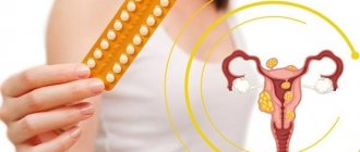
Are there any contraindications for HRT?
Yes, they exist, just like for any drug. These include:
- Vaginal bleeding of unknown origin
- Acute severe liver disease
- Acute deep vein thrombosis
- Acute thromboembolic disease, hereditary and acquired thrombophilias, previous thromboembolism or deep vein thrombosis.
- Porphyria
- Breast cancer and endometrial cancer.
In addition, there are a number of diseases for which the use of hormone replacement therapy is limited, therefore, before prescribing replacement therapy, a doctor’s consultation and examination are necessary.