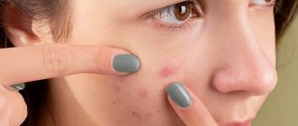
What kinds of post-acne acne occur?
There are 3 types of post-acne, not only age spots:
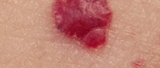
- Scars are the most undesirable complication of post-acne
- Pigment spots – brown spots on the sites of former rashes
- Congestive erythema - redness accompanied by compaction from an unresolved element
All three types of post-acne can be present on the skin either simultaneously or separately, depending on individual characteristics and the tendency to develop pigmentation or scars.
The whole truth about spider veins
Spider veins are not a disease!
So, spider veins or, as they are often called capillaries (telangiectasis), are not exactly a disease, but rather a cosmetic problem! This is the honest truth, which is often passed off as varicose veins.
Yes, on the one hand it is varicose veins, but varicose veins are at the level of skin vessels - capillaries and intradermal veins. Such microvaricose veins do not cause any painful symptoms, pain, cramps, swelling, or blood clots. Nothing. Only aesthetic discomfort.
Patients who often come in with pain in the legs, swelling, and discomfort in the calves notice spider veins on their legs and place all the blame on them. And therefore, instead of “treating” with a newfangled “anti-varicose” gel in the area of blood vessels, we recommend looking for the real cause of your discomfort in the condition of your legs (meaning symptoms) - for example, “heavy leg” syndrome, flat feet, chronic venous insufficiency, spinal osteochondrosis, etc. d.
Spider veins are not a disease, which means there is no need to strengthen the blood vessels!
Strengthening blood vessels with medications and gels is a waste of time. You can train muscles, ligaments, and develop endurance. But the blood vessels cannot be strengthened even with the help of medications.
What do we know about spider veins (webs)?
The problem is cosmetic and widespread, spider veins appear at different ages in approximately 70% of women due to the fact that female sex hormones are synthesized in the female body, starting from adolescence, and this stimulates the translucency of intradermal veins, and with age, the appearance of such unwanted elements like spider veins.
For example, a 9-month pregnancy and the accompanying changes in a woman’s body at the hormonal level, as a rule, aggravate the problem. This is normal and physiological, and most importantly, harmless.
That is, you can be calm about your health.
Is there a way to prevent the appearance of unwanted vascular elements?
You can read for hours on the Internet about preventing the appearance of ill-fated blood vessels. And even watch it on TV in popular talk shows. This is a good business - the problem is very common, and if you come up with some kind of “miracle remedy”, many will definitely try it and buy it. There used to be a drug called Asklezan - both tablets and gel. It was necessary to smear and drink 4-6 courses and they supposedly became smaller or “faded” or decreased. In general, they wrote about it in newspaper advertising, people ran and bought it. They smeared themselves. Well, of course, the effect is zero.
There is no way to prevent the appearance of spider veins!
They will gradually appear at different rates in different periods of life.
And if you don’t like it, you can think about treating existing unaesthetic telangiectasis, but it is impossible to slow down or stop this process, even if you take courses of venotonics and daily smear the area of the spider veins with various venotonic creams with or without leeches. This will not affect the process of their appearance and progression in any way.
Treatment of spider veins with medications is useless!
The basic concept of treatment for telangiectasis is not treatment of the whole body or strengthening of blood vessels or drug support by introducing any drugs into the area of spider veins using gels - this is a waste of time. Not a single star will fade, not a single one will disappear. It is a fact. All we can do is inject a sclerosant into the vascular element so that it damages the inner layer of the asterisk and it closes (sclerotherapy, or rather microsclerotherapy) or electrocoagulate the vessels every millimeter or percutaneously treat with a laser again every mm.
That is, the main concept of treatment is to destroy the vascular element in one way or another without damaging anything around it.
Percutaneous laser coagulation
Special lasers are used to treat telangiectasis. The basic principle of treatment is this: a laser beam passes through the skin and is absorbed by the red blood in the vessel, after which the blood heats up and explodes. The vessel is “brewed”. This is a very good method, but only for small and very superficially located meshes, for example, on the face and torso. On his feet he works very unsteadily and it is difficult to get results. Used in Russia mainly by cosmetologists.
In my opinion, a significant disadvantage of percutaneous laser coagulation for leg telangiectasis is the limitation in the diameter of the vessels, the high pain of the procedure and the high probability of difficult to predict side effects (burns of the skin surface, depigmentation in the affected area).
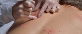
Electrocoagulation of capillaries
Using a very thin needle-electrode, many injections are made into the capillaries with simultaneous exposure to electric current. The procedure is painful and requires anesthesia with a skin anesthetic cream (EMLA on the legs takes effect after 2 hours of exposure under cling film). Therefore, you either smear yourself with Emla cream at home and wrap your legs in film and come to the doctor, or you receive hundreds of rather painful injections. The frequency of injections into the capillary is every 1-1.5 mm. That is, 1 cm of capillary will require 6-10 injections.
Sclerotherapy is the method of choice
Sclerotherapy is an injection technique for the treatment of spider veins and reticular veins. It is most effective against small veins and capillaries. Its meaning is to introduce a sclerosant into the lumen of the vessel, which causes chemical damage to the inner layer of the vessel, followed by its “closure”. After the vessel has closed, it begins to gradually dissolve (several weeks - several months).
Today, sclerotherapy of reticular (intradermal veins) and capillaries is called microsclerotherapy and has many modifications; moreover, everyone performs it differently, so the results can also be very different.
Types of microsclerotherapy
There is a technique of liquid sclerotherapy, when the sclerosant is administered in the form of a solution and this works well on small capillaries with low-intensity blood flow, and a technique of introducing foamed sclerosant (foam-form sclerotherapy), when air or other gas (CO2, for example) is added to the sclerosant, whipped by distillation through an adapter from one syringe to another and after 10-20 strokes a stable foam is obtained.
The sclerosant foam introduced into the lumen of the vessel expels blood well and is well retained there. Foam-form sclerotherapy works better on vessels with more intense blood flow.
Liquid sclerosant works more intelligently and gently, with a minimum of side effects, foam is more aggressive. In the same treatment area, liquid sclerotherapy can be used for some veins and capillaries, and foam for others. A phlebologist can use various scleropreparations, of different concentrations, in liquid or foam form during one session on one treatment area. It all depends on his experience, working style, etc. This is about the active advertising of the miracle method of foam-form sclerotherapy, which is separately positioned as a newfangled and very effective method. We've been using foam for about 10 years and it works great in certain situations, but not much else, especially with mesh.
Microsclerotherapy - a special approach
When it comes to sclerotherapy for spider veins, it is necessary to immediately clarify the situation.
This is an intelligent problem and requires a certain doctor’s experience and a certain technological approach.
It is necessary to take into account the volumes of the administered drug and its concentration. On the other hand, today microsclerotherapy is a methodologically verified procedure, and if everything is done according to technology, then the results will be as expected.
What patients don't understand
First of all, after sclerotherapy sessions, aesthetic results will not be immediate, but after some time. The fact is that closed vessels are visible through the skin and are noticeable. That is, if after the injections the spider vein does not close, then it does not change its color and remains the same as it was; if it closes, it darkens (the remaining small amount of coagulated blood shines through the skin).
The entire time that sclerotic telangiectasis is resolving, it will be noticeable. As soon as it dissolves, the marks will disappear.
This applies not only to sclerotherapy, but to any other effective treatment method. And it is important to understand this before you decide to undergo sclerotherapy. It is necessary to choose the right time for treatment so that you have a reserve of time for aesthetic rehabilitation.
How to get rid of spider veins?
It is clear that you are reading this to understand how you can improve the aesthetics of your legs. Considering the fact that in most cases the method of choice for primary treatment is sclerotherapy, it makes sense to see a phlebologist. An ultrasound of the venous system will be performed and treatment will be recommended. Naturally, in cases of small red capillaries, percutaneous laser coagulation (PLC) may be recommended to you, but this is unlikely, since PLC is usually used for follow-up treatment (only small red capillaries) and usually by cosmetologists.
In most cases of reticular varicose veins, the treatment area includes reticular (feeding) veins (blue), large and medium capillaries (purple) and small ones (red).
In order for the treatment to be effective, the phlebologist begins with the reticular (feeding) veins (marked with blue arrows in the photo), then moves on to large capillaries, then to medium and small ones. Of course, only red vessels can be treated, but practice shows that this is ineffective if the feeding reticular vein remains.
Stage one - sclerosing the “feeding” reticular veins
Often reticular veins communicating with telangiectasis are clearly visible without any equipment (magnifying glasses, polarized light). However, at the same time, poor results of sclerotherapy are associated with poor visualization of these very feeding veins, which can be obscured by the spider veins located above them. A solution to this problem has been proposed by companies that produce Veinviewers - devices based on the thermal imaging effect, which allow brilliant visualization of intradermal veins.
The phlebologist begins the session under the control of Veinviewer, punctures the feeding vein and closes it by injecting a sclerosant.
This is a simple and effective technology that we have been using for over a year to more successfully treat spider veins. Moreover, Venovisor makes it possible to clearly see the vein from which the unwanted vascular element is filled. Veinviewer technology is the key to understanding the problem of spider veins.
Stage two - sclerosing spider veins (telangiectasis)
This is a very simple manipulation at first glance - injecting the drug into the spider veins. The vascular network is punctured with a very thin needle and a small amount of sclerotherapy is injected into it. The introduction is slow and very small in volume.
This allows you to avoid long-term side reactions (matting (see below), pigmentation). Sclerotherapy can be most successful with multiple injections and small amounts of sclerotherapy.
After the introduction of sclerosant, we observe the redness of the element and its reaction. There may be a slight tingling sensation in the injection area. Thus, in the first session, as a rule, the feeding reticular veins are treated, in the second and subsequent sessions - telangiectasis (spider veins).
A few words about haste in treatment
You come 2-3 months before your vacation with the desire to get rid of capillaries, but you did not know that you cannot rush in the treatment of spider veins.
A number of side effects, which can then subside for months and years, are associated not only with the sclerotherapy technique, but also with short breaks between sessions.
Ideally, a session on one leg in the same zone should be carried out at intervals of 3-4 weeks. This is the optimal schedule. That is why we recommend treating spider veins as planned, taking into account your vacations and beach holidays and “much in advance.”
How to make injections painless and improve the results of sclerotherapy?
In microsclerotherapy, injections are performed with cosmetic needles with a diameter of 0.3 mm. These are very thin needles and the injection is insensitive. However, some patients are afraid of injections and for them this type of treatment creates psychological stress. We have solved this problem because we perform cryosclerotherapy. The technique was born in the USA and, with the light hand of the well-known Luis Navarro, was called painless sclerotherapy.
This new method involves cooling the skin for a few seconds with a blast of cold air before injection, creating cold anesthesia of the skin. This is enough so that the patient does not feel the injection.
The miracle of cryosclerotherapy
As it turned out later, this is not the only positive thing from cryosclerotherapy - bruises, hematomas, pigmentation and other side effects after the session are much less than after conventional sclerotherapy. This is due to local vasospasm in the treatment area. And, as a result, in spasmodic vessels the sclerosant works more effectively with a lower concentration and a smaller injected volume.
Compression and sclerotherapy
It has always been believed that round-the-clock compression is necessary for the entire period of sclerotherapy - during the day with stockings, at night with an elastic bandage, plus another 2 months after the end of treatment. That is, only six months. For many patients this was unbearable. But if this is true, then only for sclerosis of large veins. When performing microsclerotherapy of spider veins and reticular veins, compression is usually needed until the evening - either a bandage or stocking. The main purpose of compression in such cases is to reduce the formation of bruises after the session. In rare cases, the daily compression regimen can be extended to 3-5 days.
Modern technology for microsclerotherapy allows this type of treatment to be carried out with virtually no compression and at any time of the year, even in summer.
Innovations in sclerotherapy
Globally, the microsclerotherapy technique has not changed. Same drugs, same injections. Foam-form sclerotherapy, when used on spider veins, usually does not live up to expectations, so sclerotherapy with a liquid solution is preferable. However, since 2013 we have introduced a couple of innovations in technology: cooling the skin during the procedure and the use of a Veinviewer.
These improvements make the procedure more comfortable, reduce the number of side effects, and improve control over the administration of the drug.
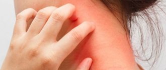
Possible side effects of microsclerotherapy
Pigmentation
The most common side effect of microsclerotherapy is pigmentation at the site of sclerotic veins and spider veins. After administration of the sclerosant, the inner layer of the vessel is damaged; at first the vessel disappears, but after a few minutes it fills with blood again. If the vessel subsequently thromboses (closes), then a black tar-like mass (coagulum) is formed in it, which is darker than blood and the vessel shines through the skin brighter than the original one. Over time, the closed vessel with coagulum dissolves. Usually this is several months. If this process is long and the coagula is quite pronounced, then iron is deposited in the skin and it becomes light brown. Actually this is called pigmentation. In most cases, pigmentation goes away within six months, but in 5% of cases it can take a year and in 1% of cases up to 2 years. But it always passes, which makes me happy.
The severity and duration of pigmentation after sclerotherapy is individual and depends on the characteristics of pigment metabolism in the patient’s body.
Matting (neovascularization)
Matting is a reaction in the form of the appearance of small capillaries in the treatment area. This is a complex and sometimes difficult to explain and predictable phenomenon. It is largely determined by hormonal levels (manifested in women), as well as by the individual reaction to the administration of sclerosant. Most often associated with the use of high concentrations of scleropreparation or a large volume of administration.
That is why you should not try to inject all the vessels at once in the first session - the dose may be large and this will cause an undesirable reaction.
Most often, matting occurs if sessions on one leg occur very often (once a week, for example). A more gradual style of treatment with longer breaks produces better results. You should also not conduct more than 3 sessions in a row on one leg without a long break (3-6 months).
How to minimize the risk of side effects during sclerotherapy?
Introduce a small amount of sclerosant through multiple injections, take long breaks, and take a long rest after 2-4 sessions. Sclerotherapy should be like the leisurely work of an artist who gradually draws the details of a picture.
That is why it is necessary to approach your treatment without haste and without any limiting circumstances. Choose a time and go to the procedures calmly.
#FORM_PSK1#
The main predisposing factors for the appearance of post-acne
- Severity of acne – acne of III and IV severity often leads to complications, because affect the deep layers of the skin.
- Individual skin characteristics, genetic predisposition - such as dark skin color, a tendency to increase melanin production at the slightest inflammation or damage to the skin.
- Lack of treatment, inappropriate treatment, or late treatment is when the process has been present in the skin for a long time and has affected deep tissues.
- Skin injuries during mechanical cleaning, i.e. when roughly squeezing out pimples.
The presence of at least one predisposing factor may be the cause of the development of post-acne.
Heparin ointment
Heparin ointment is a combination drug for external use, which contains three pharmacologically active substances: heparin, benzocaine and benzylbenzocaine. It is unlikely that the latter two will object that the main character of the drug, which determines its entire pharmacological “policy,” is heparin. This direct-acting anticoagulant is capable of keeping the inflammatory process in check and, most importantly, providing an antithrombotic effect, the mechanism of which (or rather its description) is not very simple for the ignorant. Heparin binds to antithrombin III (a specific protein of the blood coagulation system), changing the spatial arrangement of atoms in its molecule and promoting the formation of antithrombin III + serine protease complexes of the coagulation system. The result of these disturbances is the blocking of thrombin, suppression of the enzymatic activity of plasma coagulation factors, plasmin and kallikrein. The thrombin binding reaction has an electrostatic basis and is largely determined by the length of the heparin molecule (only a small section of it is able to bind to antithrombin III, in fact, it is responsible for the anticoagulation properties of the entire molecule). At the end of the thrombin inhibition reaction, heparin is released from the “embrace” of antithrombin III and is again ready to work and protect the body from the consequences of blood thickening: it reduces its viscosity, reduces the permeability of the vascular walls stimulated by bradykinin, histamine and other inflammatory mediators, thereby preventing blood stagnation. Heparin is able to settle on the membranes of endothelium, platelets, erythrocytes and leukocytes, preventing their adhesion and aggregation. It inhibits the activity of the enzyme lipoprotein lipase, which is one of the regulators of lipid metabolism, thereby slowing down the development of atherosclerosis. Heparin also has, to a certain extent, an antiallergic effect, which is achieved by binding individual components of the complement system, which creates an obstacle to the cooperation of lymphocytes and suppresses the synthesis of immunoglobulins.
Another component of heparin ointment - benzylbenzocaine (benzyl ester of nicotinic acid) - increases the lumen of superficial vessels, thereby creating favorable conditions for the absorption of heparin from the surface of the skin. And the last member of the antithrombotic trio called “heparin ointment” is the local anesthetic benzocaine. It prevents the penetration of sodium ions through cell membranes, “pushes out” calcium ions from membrane receptors, blocks the generation and conduction of impulses along nerve fibers, which leads to a decrease in the severity of pain. When applied to the skin, it is practically not absorbed, thus not having an undesirable systemic effect on the body. It begins to act within 1 minute from the moment of application, maintaining activity for 15–20 minutes.
Heparin ointment, as trivial as it may sound, is available in the form of an ointment. It should be applied in a thin layer at a rate of 0.5-1 g to the affected area with a diameter of 3-5 cm, and then gently rubbed into the skin. This manipulation is carried out regularly 2-3 times a day until the signs of inflammation completely disappear. On average, this takes from 3 to 7 days. Longer therapeutic courses using heparin ointment can only be carried out in consultation with a doctor. For external hemorrhoidal thrombosis, rectal tampons are used: the ointment is applied to a linen or calico pad, which is then applied directly to the affected nodes and fixed. The course of treatment, subject to daily use of the ointment, ranges from 3 to 14 days. And in conclusion, important information about the features of combining heparin ointment with other drugs: it should not be used in conjunction with non-steroidal anti-inflammatory drugs, tetracycline antibiotics and antihistamines.
How to avoid age spots and scars
- Start acne treatment immediately. Even mild forms cannot be ignored - grade 0-2 acne, when there are single elements on the skin.
- Don't push! A banal but difficult point to implement. Skin cleansing can be done and sometimes even necessary, but it should be done carefully and competently by a cosmetologist.
- Use medications with anti-inflammatory effects. Not to be confused with antibacterial! Anti-inflammatory drugs have both preventive and therapeutic effects.
Recommendations follow from the previous paragraph - exclusion of predisposing factors.
Heparin
Heparin is a direct anticoagulant that suppresses the activity of the blood coagulation system and prevents the formation of blood clots. Once in the blood, it activates the main protein of the blood coagulation system - antithrombin III, as a result of which the rate of onset of its anticoagulant effect increases. It prevents the conversion of prothrombin to thrombin, suppresses the activity of the latter (and in addition to this, activated factor X), and reduces the ability of platelets to aggregate (stick together). Stimulates blood circulation in the kidneys, increases the resistance of cerebral vessels, and has a hypolipidemic effect. Reduces the activity of pulmonary surfactant, inhibits the excessive formation of aldosterone by the adrenal cortex, and activates the hormone of the parathyroid glands. According to some data, it has immunosuppressive activity. In patients with coronary heart disease (in combination with acetylsalicylic acid) it reduces the risk of acute coronary thrombosis, myocardial infarction and sudden coronary death. Reduces the number of relapses of myocardial infarction and mortality in post-infarction patients. High doses of heparin help with pulmonary embolism and acute venous thrombosis, low doses “work well” as part of preventive measures to prevent venous thromboembolism, including after surgery. With intravenous administration of heparin, blood clotting slows down almost immediately, with intramuscular administration - after 15-30 minutes, with subcutaneous administration - after 20-60 minutes. The duration of the anticoagulant effect is, respectively, 4-5 hours with intravenous, 6 hours with intramuscular and 8 hours with subcutaneous administration. At the same time, the main therapeutic effect of heparin - the prevention of blood clots - lasts much longer. An insufficient amount of antithrombin III in the blood or directly at the site of thrombosis can neutralize the antithrombotic effect of the drug. When applied externally (and the drug with the trade name “heparin” is also available in the form of a gel) it has antithrombotic, antiexudative and mild anti-inflammatory effects.
Prevents the formation of thrombin, suppresses the activity of hyaluronidase, enhances the fibrinolytic properties of blood. Heparin, which penetrates through the skin, relieves inflammation, improves blood circulation in the microcirculation, stimulates tissue metabolism, which ensures accelerated resorption of hematomas and blood clots.
The use of anticoagulants in various pathological conditions (including acute and emergency) is of persistent interest both within clinical practice and from the standpoint of improving the quality of medical care. Unfractionated heparin, which is discussed in this article, is most often used to prevent and treat thrombotic disorders, including deep vein thrombosis and thromboembolism (acute blockage of a blood vessel by a blood clot). Statistics show that approximately 50% of patients with acute venous thrombosis receive heparin as anticoagulant therapy. Since heparin has a fairly short therapeutic window (the difference between the minimum toxic and minimum effective concentrations of the drug) and can cause various types of hemorrhagic complications, the patient's condition must be carefully monitored when using it. In the list of drugs published by the American Institute for Healthcare Improvement, the misuse of which has led to serious consequences, heparin ranks third. Another highly respected institution, the US Institute for Drug Safety, has classified heparin as a drug whose use requires special precautions. In the United States, there is a program in which health organizations provide information about errors in drug therapy and side effects of drugs. So, the largest number of such mistakes are made when using anticoagulants, incl. and heparin. In this regard, heparin in the form of solutions should be used only in hospital settings.
How to get rid of post-acne
Recommendations of the International Society of Dermatologists and Cosmetologists: 1st line drugs for acne and post-acne treatment are retinoids .
- Have an anti-inflammatory effect;
- Normalize the functioning of pigment cells, lighten pigment spots;
- Normalizes the functioning of the sebaceous glands. With acne, the amount and composition of sebum changes - the main reason for the development of acne;
- Increase local immunity, which helps fight inflammation;
- Stimulate the synthesis of collagen necessary to fight scars.
Due to such a wide range of cosmetic effects on the skin, retinoids are recognized as the gold standard for the treatment of acne and post-acne.
The result of using retinoids:
- lightening pigment spots
- smoothing skin texture
- reduction of scar depth
Retinol benefits:
- Physiological – is a substance familiar to the skin, because the body itself supplies vitamin A to the skin in low concentrations;
- Has no toxic effect;
- Suppresses the increased activity of melanocytes, without destroying them, but normalizes melanin production;
- Penetrates well into the skin, reaching the necessary layers;
- Stimulates cellular renewal, preventing colored cells from remaining on the surface, and accelerates their exfoliation.
Indications for use
Since heparin ointment is available without a prescription, you need to know what it is used for. Common pathologies for which the drug is effective are the following:
- Chronic hemorrhoids.
- Inflammation of hemorrhoids after childbirth.
- Lymphatic failure.
- Thrombophlebitis.
- Superficial mastitis.
In addition, the product allows you to quickly eliminate the consequences of injuries and bruises of muscle tissue, joints, tendons, etc. It can be used to prevent varicose veins. It is also effective in the early stages of the development of this pathology, as it relieves pain and heaviness in the legs, normalizes microcirculation and promotes the resorption of formed blood clots.
How to use heparin ointment
It is important to know heparin ointment, what it is intended for and how to use it for various pathologies. Only if you follow the rules of use will you be able to achieve the desired effect in the shortest possible time. The product is applied to the affected area in a thin layer until the inflammatory symptoms disappear completely. Approximately 0.5-1 g or 2-4 cm is needed for an area with a diameter of 3-5 cm. On average, it takes 3 to 7 days for a positive result. A longer course of treatment must be agreed with the doctor.
In the early stages of varicose veins development, with the appearance of spider veins and slight expansion, the medicine is applied to areas where there are altered vessels. Manipulations are carried out for two weeks 3 times a day.
Heparin ointment, the instructions emphasize this, is an effective remedy for treating bruises. Due to the anticoagulant properties of the main active ingredient, the drug accelerates the resorption of blood clots. Other active components in the ointment provide a vasodilator and local analgesic effect. To eliminate bruises that are the result of severe contusions, the product is applied to the damaged area and rubbed in with gentle massage movements. Manipulations are performed 2-3 times a day for 5-15 days. But according to reviews, subcutaneous hematomas can be eliminated in 2-4 days in this way.
Heparin ointment against hemorrhoids, instructions for use indicate this, is used as follows:
- A small amount of the product is applied to a cotton pad.
- It is applied to inflamed nodes.
- When internal cones form, the drug is administered using a special nozzle.
As a rule, these manipulations are carried out until the symptoms of hemorrhoids disappear. Usually 5-14 days are enough for this.