Kidney stones (Nephrolithiasis, Kidney stone disease)
Conservative treatment
Treatment of nephrolithiasis can be conservative or surgical and in all cases is aimed at removing kidney stones, eliminating infection and preventing the re-formation of stones.
For small kidney stones (up to 3 mm), which can be passed independently, plenty of water load and a diet excluding meat and offal are prescribed. For urate stones, a dairy-vegetable diet that alkalizes urine and alkaline mineral waters (Borjomi, Essentuki) is recommended; for phosphate stones - take acidic mineral waters (Kislovodsk, Zheleznovodsk, Truskavets), etc. Additionally, under the supervision of a urologist, medications that dissolve kidney stones can be used (for example, citrate therapy for urate stones).
First aid for renal colic
With the development of renal colic, therapeutic measures are aimed at relieving obstruction and pain. For this purpose, injections of platyphylline, metamizole sodium, morphine or combined analgesics in combination with atropine solution are used; A warm sitz bath is performed and a heating pad is applied to the lumbar region. In case of intractable renal colic, novocaine blockade of the spermatic cord (in men) or round ligament of the uterus (in women) and catheterization of the ureter are required.
Surgery
Surgical removal of stones is indicated for frequent renal colic, secondary pyelonephritis, large stones, ureteral strictures, hydronephrosis, kidney blockade, threatening hematuria, stones in a single kidney, coral stones. For nephrolithiasis, remote lithotripsy is used to avoid any intervention in the body and remove stone fragments through the urinary tract. For stones up to 2 cm in diameter, you can use the “flexible retrograde nephrolithotripsy” method, as well as percutaneous nephrolitholapaxy, which allows you to remove the stone through a puncture in the kidney.
Open or laparoscopic interventions to remove stones - pyelolithotomy (dissection of the pelvis) and nephrolithotomy (dissection of the parenchyma) are rarely resorted to, mainly when minimally invasive surgery is ineffective. In case of complicated kidney stone disease and loss of kidney function, nephrectomy is indicated. After removal of stones, patients are recommended spa treatment, lifelong diet, and elimination of associated risk factors.
Why are they formed?
- Insufficient amount of urine.
With a small volume of urine, stones may occur. This occurs due to dehydration, hot weather or low water consumption. Due to the small volume of urine, it becomes concentrated and dark in color. For prevention, doctors advise drinking a lot of water, especially when working or in hot weather.
- Poor nutrition
. A common cause of stone formation is excess calcium. And it’s not just about nutrition, it depends on how the body processes it. The second reason will be the influence of oxalic acid. It is found in food products: spinach, sorrel, parsley, nuts, cocoa. But the most dangerous are deposits of magnesium and iron salts. They form sharp ends and can damage the walls of organs.
- Gastrointestinal diseases.
Some stomach and intestinal diseases can lead to dehydration.
- Genetic diseases. Rare diseases that are inherited can cause disturbances in salt metabolism.
Symptoms
Sometimes the disease passes without symptoms, and pain appears during urination. But renal colic often manifests itself. The location of the stones may vary, so pain may occur in different places.
- The appearance of blood in the urine
. The amount of blood may vary and be insignificant, or may turn into bleeding. This is caused by injuries to the soft tissue of the kidneys and adrenal glands. - Urinary disorders.
They occur when stones pass through the urethra and bladder. Sometimes a stone can completely or partially block the passage, which leads to a complete absence of urine. Such problems can lead to pyelonephritis.
Symptoms of ICD
Signs of urolithiasis vary depending on the location of the stone:
In the kidney
The patient feels acute pain in the lower back; when moving and the stone increases in size, the condition worsens - the inner surface of the kidney cavities is injured. Dull pain in the lower back and blood in the urine appear, especially after physical exertion. The surface of the stone can serve as a favorable environment for infection; secondary chronic pyelonephritis occurs, which is manifested by chronic pain in the lumbar region, an increase in body temperature from low-grade to fever during an exacerbation. Lack of adequate treatment can lead to the complete loss of the kidney's functional abilities and the development of renal failure.
If spontaneous passage of kidney stones does not occur, acute inflammation of the kidney (pyelonephritis) may occur, which requires kidney surgery; this condition is potentially life-threatening, and the timing of acute pyelonephritis varies from person to person.
In the ureter
Acute pain that appears in the lower back and moves to the groin area, often radiating to the thigh, often to the genitals. When the stone is located in the lower region of the ureter, there is a frequent urge to urinate. When a stone completely blocks the ureter, a person feels especially acute pain in the lower back, transmitted to the abdominal area.
Often accompanied by fever, nausea, and vomiting. The common name for this condition is renal colic.
In the bladder
The pain is concentrated in the lower abdomen. When moving or urinating, the discomfort intensifies. The urge to go to the toilet is especially pronounced during physical activities. When urinating, the flow of urine may stop for a while and resume only when the person changes body position.
Why kidney stones form
Urolithiasis can be a consequence of both local and general processes in the body. The development of urolithiasis in men and women is a consequence of changes occurring in the body for reasons such as:
- excess calcium compounds in the diet;
- bacterial infection;
- long-term use of certain medications (sulfonamides, antacids, etc.);
- improper metabolism of vitamins D and A.
- Insufficient fluid intake
- Increased protein intake
Local processes include such pathologies of the genitourinary system as:
- prolonged use of a catheter;
- poor blood supply to organs;
- spinal cord injury, as a result of which the outflow of urine is disrupted; a mobile state of the kidney or, in other words, nephroptosis, leading to its displacement relative to the correct position, other pathologies - hydronephrosis, polycystic disease;
- consequences of operations on the abdominal cavity and pelvic area, tumor processes;
- cystitis, pyelonephritis, prostatitis
Formation of kidney stones
Hereditary or acquired diseases upset the balance of substances in the body, which increases the concentration of minerals that accumulate in the kidney tubules. Stagnation of urine, narrowing in the renal collecting system and limitation of physical activity are also conditions for the formation of stones. The human body has mechanisms for expelling small stones, however, with a combination of unfavorable factors (the size of the stone, its surface, weakening of the body due to stress and concomitant pathology), the stone-expelling properties fail, the stone becomes fixed in the urinary tract, begins to increase in size and create obstacles to normal urine outflow
It is not uncommon for the formation of stones to be affected by diseases such as prostatitis and prostate adenoma.
To assess the chemical composition of the stone, the Miracle Doctor clinic uses the infrared spectroscopy
. The chosen method of treating urolithiasis depends on the composition of the stone and preventive measures are prescribed. For evaluation, either all or part of the released stone is required.
Classification of stones
- Uric acid (urate) - smooth brownish formations that are formed by salts of uric acid.
- oxalate - brown, rough formations, sometimes with spikes, formed by salts of oxalic acid.
- phosphate - stones with a soft and fragile structure, grayish tint.
- mixed - the core and shell are formed from different types of salts.
- cystine - are the hardest, the surface is smooth, without thorns.
A mixed composition of stones is often found.
Diagnosis of ICD
Diagnosis of urolithiasis usually includes assessment of general clinical signs, results of laboratory and hardware tests.
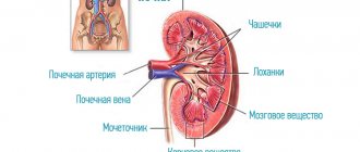
Ultrasound examination of organs
urinary ultrasound of the kidneys and ureters, bladder, parathyroid glands of the kidneys, the specialist determines the shape and contour of the organs, density, urostasis - expansion of the cavities of the kidney, the state of the collecting system.
X-ray research methods
Computed tomography with and without contrast, survey or excretory urography - these types of diagnostics of urolithiasis provide information on the number, size and location of stones, patency of the urinary tract. The required amount of research is determined by a urologist.
Laboratory methods
Laboratory tests when detecting urolithiasis make it possible to determine the possible causes of stone formation - bacteriological culture of urine, examination of the level of uric acid, ionized calcium, blood potassium and other indicators. If there is a stone passed in the urine or removed during surgery, it is very important to examine its composition in order to determine further management tactics, selection of diet, and prescription of medications.
Treatment methods for urolithiasis
Treatment of urolithiasis is mainly surgical - an operation is performed to remove the stones. If the stones are formed by uric acid salts, their dissolution is possible. In this case, a course of special medications is prescribed.
However, if the conditions that caused the stone formation remain the same, then the stones may appear again after they are removed. To avoid this, a course of preventive treatment is carried out, including adherence to a special diet, medication, maintaining the necessary water balance, and physical therapy.
By contacting JSC “Family Doctor” with complaints about a disease of the urinary organs, you will receive advice from qualified urologists. Their experience, as well as equipping the Family Doctor clinics with modern equipment, including its own laboratory, will allow you to quickly and accurately establish a diagnosis and prescribe effective treatment. Stone removal is carried out in the Hospital.
Removing Stones
Currently, stone removal is usually done using minimally invasive methods. Laparoscopy and open surgery should be resorted to only in extreme cases. First of all, methods such as pyelolithotripsy are used (stones are destroyed as a result of mechanical, ultrasound or laser action using instruments placed along the natural urinary tract) and percutaneous nephrolitholapaxy (removal is performed through an incision in the lumbar region. This is how large and coral-shaped stones are removed).
Make an appointment Do not self-medicate. Contact our specialists who will correctly diagnose and prescribe treatment.
The most important thing for the patient
- Currently, medicine still does not allow us to absolutely accurately establish the mechanism of stone formation. As a result, there are no generally accepted and highly effective methods of prevention and drug treatment for most forms of this disease.
- Surgical treatment of urolithiasis has made a tremendous leap forward in recent decades, changing beyond recognition. Today there is practically no place for open (“abdominal”) surgery.
- The main modern methods of treating urolithiasis: remote lithotripsy (crushing stones with a shock wave), contact and percutaneous lithotripsy (using modern, including flexible endoscopes and various methods of stone destruction: laser, ultrasound, pneumatic), laparoscopy.
- Unfortunately, there is no universal way to destroy stones. Remote lithotripsy is truly the most minimally invasive (minimally traumatic) method. However, it has quite a lot of limitations. In some cases, this approach is not effective at all. And it often happens that after extracorporeal lithotripsy (even in the most skillful hands), in order to finally achieve the goal (or combat complications), the additional use of other methods of crushing stones is required.
- Optimal conditions for effective and safe treatment of urolithiasis can ONLY be created in a hospital that has ALL modern methods of stone destruction and appropriate qualified specialists.
- Limitation in the choice of method of surgical treatment of urolithiasis often leads to a significant “complication of life” for both the patient and the doctor.
Every year at the Clinic, hundreds of patients from different regions of Russia receive assistance in the treatment of urolithiasis in accordance with the latest international standards. You can read the stories of patients treated at the Clinic on the page
Video reviews
Medicines and substances that increase risk
- Acetazolamide
- Efilrin
- Phosphate-binding anatcides
- Uricosuric substances
- Calcium/Vitamin D
- Guaifenesin
- Topiramate
- Triamterene
- Vitamin C
In modern medicine there is no remedy that cures urolithiasis. After pain relief and stone removal, the only goal is to prevent relapse
The process of stone formation can be controlled. Proper nutrition and plenty of water in the diet will reduce the likelihood of recurrence.
Why do kidney stones form?
There are various theories about the formation of kidney stones. The main factor contributing to the formation of stones is the excess concentration of substances (salts) in the urine from which they can form (calcium compounds, oxalates, phosphates, uric acid, etc.). Due to excess concentration, these substances form crystals and may precipitate. Fortunately, this does not normally happen due to the presence of stone formation inhibitors in the urine. Kidney stones are mainly formed when the urine is highly saturated with salts or lacks inhibitors, thus crystals first form in the urine, which then form stones.
The composition and shape of stones vary depending on the substance from which they are formed. The figure below shows the main types of stones in the urinary system
Factors contributing to stone formation
1. Consumption of foods rich in stone-forming substances
2. Diseases and conditions associated with stone formation
- Hyperparathyroidism
- Renal tubular acidosis
- Jejunoileal anastomosis
- Crohn's disease
- Condition after ileal resection
- Malabsorption syndrome
- Sarcoidosis
- Hyperthyroidism
3. Family history of urolithiasis
4. Drugs associated with stone formation
5. Anomalies in the structure of the urinary system associated with stone formation
- Tubular ectasia
- LMS stricture
- Diverticulum/calyx cyst
- Ureteral stricture
- Horseshoe kidney
- Urinary tract infections