It is under this name “thyroid hyperplasia” that endocrinologists with extensive experience diagnose patients. In medical terminology, this concept is outdated; the term “goiter” is more often used. Patients with goiter live in areas where the soil and groundwater contain little iodine salts.
Due to iodine deficiency, pathological hypertrophy of the thyroid gland develops in the body, accompanied by functional disorders. Most often, this disease occurs in the countries of the Eastern Mediterranean: Israel, Syria, etc.
Causes of hyperplasia
How hyperplasia appears in the body is not known for certain. However, it is known that a number of reasons contribute to the development of this pathological process and, above all, the problem of the secretion of prohormones and their conversion into hormones. It is important to remember that hyperplasia is not a specific diagnosis, but a syndrome that requires clarification of etiology, histology, etc. So, the factors for the progress of hyperplasia are as follows:
- deficiency of iodine salts in the body;
- pathogens;
- autoimmune thyroiditis;
- immune pathologies;
- pregnancy process;
- neoplasms in the pituitary gland and hypothalamus;
- malignant neoplasms in the thyroid gland;
- congenital pathologies of the thyroid gland;
- taking medications that disrupt the functioning of the thyroid gland.
The main reason is iodine deficiency in the environment. It was assumed that iodized salt would solve the problem once and for all, but still a third of the world's people suffer from a lack of iodine in the body.
How to recognize hyperplasia?
The surest way to find out whether you are really sick or not is to make an appointment with a doctor who has enough ways to prove or disprove the diagnosis.
If you know that you are susceptible to such diseases due to genetics, or you are susceptible to certain risk factors, then you need to monitor the following symptoms:
- severe nervous excitability;
- increased nervousness, unhealthy shine in the eyes;
- dilated pupils;
- incomprehensible losses and gains of extra pounds. For example, in one week a patient may lose 10 kilograms of weight, and the next week these kilograms will return in an incomprehensible way, but the diet will not be changed in any way;
- swelling in the neck area.
If you feel something like this, you need to make an appointment with a doctor as soon as possible. Even if the problem is not the thyroid gland or an increase in its size, the disease is in any case related to hormones - and this is what the endocrinologist
. When you receive a referral to him, then you can go to him with a pure soul and complain about everything that bothers you.
Classification of gland hyperplasia
The dimensions of the thyroid gland are given by endocrinologists to 3 degrees of goiter:
- no goiter;
- hyperplasia is present, but not visually displayed;
- the size of the gland is increased up to a change in the shape of the neck.
Some endocrinologists still use the old classification system, which distinguishes six degrees of hyperplasia:
- zero degree, in which the thyroid gland cannot be palpated during palpation;
- the first degree, when the thyroid gland itself cannot be palpated, is noted only when swallowing hyperplasia of the isthmus of the thyroid gland;
- second degree - the entire thyroid gland can be felt when swallowing;
- third degree is characterized by mild neck deformity due to thyroid hyperplasia;
- fourth degree, in which severe deformation of the neck is observed;
- the last, fifth degree, when an overgrown goiter compresses neighboring organs and tissues due to its enormous size.
Forms of hyperplasia
The structure of an enlarged thyroid gland differs in the following forms:
- Diffuse hyperplasia of the thyroid gland. All thyroid tissue increases evenly. Typically, hyperplasia syndrome accompanies diseases such as de Kerwin's and Riedel's thyroiditis, autoimmune, diffuse toxic goiter, and tumors of a hormonal nature in other organs.
- Nodal type increase. Nodular hyperplasia of the thyroid gland is based on multiple or single nodes that accumulate to form compactions.
- Diffuse nodular hyperplasia of the thyroid gland, when nodes are palpated with uniform hypertrophy of the gland.
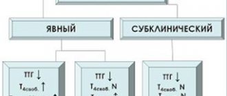
Stages of the natural course of iodine deficiency goiter
Nodular hyperplasia is considered the most dangerous of all forms. This type of enlargement requires a fine-needle biopsy to rule out malignancy. An enlarged thyroid gland can involve the entire gland or individual parts of it, symmetrically disrupting the functioning of other glands. Thus, with hyperplasia of the right lobe of the thyroid gland, a malfunction of the right mammary gland in women is detected.
Symptoms of hyperplasia
The manifestation of hyperplasia depends on the degree and type of disruption of the gland. Some disorders do not make themselves manifest clinically; only a deformation of the neck may indicate an ongoing pathology. In the initial stages of development, many diseases are also asymptomatic. If they are not treated promptly, the gland enlarges so much that difficulty breathing and swallowing occurs.
Pronounced hyperplasia
Pronounced symptoms usually appear in elderly patients with untimely treatment of the disease. Here they are:
- difficulties with swallowing due to compression of the esophagus by a hypertrophied gland, which makes it difficult for the patient to swallow even a pill on his own;
- breathing difficulties that occur while lying down or during physical activity due to the pressure of the hypertrophied gland on the trachea. Patients feel short of breath;
- The timbre of the voice changes due to compression of the nerve innervating the vocal cords. Usually the voice becomes lower and sometimes becomes hoarse. The symptom is most pronounced in speakers;
- cough;
- the appearance of circulatory disorders due to compression of blood vessels. There is redness of the neck and face;
- pain associated with bleeding due to injury to blood vessels
With nodular hyperplasia of the thyroid gland, a number of other symptoms appear: insomnia, body weight problems, hypertension, arrhythmia, increased panic, irritability, depression.
It is necessary to determine hyperplasia of the thyroid gland in children taking into account the characteristics of the child’s growth rate, his age, relationships with peers, and the dynamics of school performance.
The level of thyrotropin, normal for this age, but high for an adult, can cause misunderstandings in the relationship between “fathers” and “children”.
Hypothyroidism in an adult does not mean a similar syndrome in a child. In any case, you will need to consult an endocrinologist.
What causes hyperplasia?
Enlargement of the thyroid gland is a completely natural, although painful process, which is caused by certain changes in the condition of the body. Simply put, the thyroid gland will not simply enlarge. This outcome can be caused, for example, by a lack of iodine in the body or a hormonal imbalance. Receiving messages from the body that some hormone is missing, the gland begins to actively produce it, and itself increases due to active cell division. This is what is called hyperplasia.
Treatment of hyperplasia
The purpose of the treatment regimen is selected after a detailed diagnosis and clarification of the cause of the disease. What distinguishes thyroid hyperplasia from other diseases? First of all, treatment using conservative methods, without the use of surgical intervention. For small or medium enlargements of the thyroid gland, hormonal medications in the form of tablets are prescribed.
Treatment with hormones for gland hypertrophy compensates for the lack of hormones in the body. If it is possible to restore the normal natural production of thyroid hormones, then the growth of the gland stops and gradually the volume of the gland returns to normal. If it is impossible to achieve the desired effect with replacement therapy, the thyroid gland is removed surgically.
The thyroid gland is also surgically removed in case of significant enlargement, when it begins to compress the trachea and esophagus and causes difficulty breathing and swallowing. If X-rays and computed tomography of the neck show such compression of organs, there is no doubt about surgical intervention.
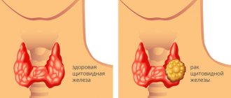
The next reason for surgical removal of the thyroid gland is malignant neoplasms. One in twenty thyroid tumors is malignant.
Although there is no indication for surgery for a cosmetic defect, some patients still require thyroidectomy to eliminate the visible effect of hyperplasia.
Adenomatous hyperplasia of the thyroid gland
Sometimes an isotope of radioactive iodine is used as treatment, which makes it possible to monitor growth processes in the thyroid gland. This method allows you to reduce the volume of the gland. A prerequisite after the administration of radioactive iodine is the use of synthetic drugs containing thyroid hormones.
When the size of the gland can be reduced without the use of hormonal therapy and, especially, surgical intervention, they are limited to adjusting the diet to include seafood and iodine-containing foods.
Note that the treatment regimen for hyperplasia of the right lobe of the thyroid gland does not differ in the increase in the entire gland or another lobe.
Discussion
Currently, the dominant theory of pituitary origin is that the vast majority of pituitary adenomas arise as a result of gene disorders in a single cell.
Subsequently, its tumor transformation occurs, followed by clonal expansion and adenoma formation. Most TSH-AGs are plurihormonal tumors that originate from pituitary stem cells and have the potential to differentiate in various directions. In 30-40% of cases, TSH-AH is characterized by hypersecretion of other pituitary hormones - most often GH and PRL, less often - gonadotropins (LH/FSH). Even in the case of hypersecretion of only TSH, in most TSH-AGs, genetic analysis reveals the expression of the GH and PRL genes [17, 18]. Hypersecretion of TSH by tumor tissue leads to hyperstimulation of the thyroid gland and, as a consequence, hypersecretion of thyroid hormone. T4 and St. T3 [11]. This is the so-called “central hyperthyroidism”. Clinically, it is manifested by symptoms of hyperthyroidism, and with a significant tumor size, also by signs of tumor mass effect: visual and neurological disorders. Violation of hypothalamic regulation is also an important link in the oncogenesis of the adenohypophysis. Thus, chronic hypofunction of the peripheral endocrine glands induces constant hypothalamic stimulation with subsequent hyperplasia and the formation of a pituitary tumor [9, 13, 19-22]. It is well known that with manifest PG the concentration of TSH in the blood is increased, and st. T4 is reduced. In such patients who do not receive thyroid hormone therapy for a long time, hyperplasia of thyroid-trophic cells may develop as a result of the lack of the inhibitory effect of these hormones on the hypothalamus. This in turn leads to an increase in the production of TRH (thyrotropin-releasing hormone), which stimulates the pituitary gland, which is manifested by the development of hyperplasia of thyrotrophic cells. Cases of compression of the optic nerves by the hyperplastic pituitary gland and the appearance of neuro-ophthalmological symptoms have been described [cit. according to 7].
Increased secretion of TRH by the hypothalamus can lead not only to hyperplasia of thyrotrophic cells and increased secretion of TSH, but also to hyperplasia of lactotrophic cells and increased synthesis and secretion of PRL, which is accompanied by an increase in the level of PRL in the blood and clinical manifestations of hyperprolactinemia [23]. Hyperplasia of thyroid cells as a result of PG is most common in women and is usually reversible with the use of adequate doses of thyroid hormones, which is manifested by normalization of TSH levels, st. T4 and St. T3 and also PRL.
However, long-term stimulation of TRH can lead to the formation of so-called “secondary” TSH-AG [10]. In a study by N. Ryan [9] of autopsy material from 64 pituitary glands from patients with chronic hypothyroidism, thyrotrophic hyperplasia was found in most cases. Diffuse hyperplasia of thyrotrophic cells was detected in 69% of samples, and nodular hyperplasia of thyrotrophic cells was detected in 25%. In 12% of cases, the changes were regarded as an intermediate stage between nodular hyperplasia and microadenoma. Five pituitary microadenomas with positive TSH expression were identified. Lactotrophic hyperplasia was present in 20% of samples.
Differential diagnosis (including MRI signs) of adenohypophysis hyperplasia and true hypertension against the background of PG is often difficult, and as a result, difficulties arise in choosing a treatment method, which is fundamentally different for these types of pathologies. Thus, for thyrotrophic hyperplasia against the background of PG, an effective treatment method is therapy with L-T4 drugs, which leads to normalization of the level of thyroid hormones and, as a consequence, a decrease in the level of TSH and regression of thyrotrophic hyperplasia, which is manifested by a decrease in volume in the chiasmatic-sellar region. The duration from the start of treatment is usually 1-4 months, although in some cases regression of formation in the chiasmal-sellar region can be observed during the 1st week of L-T4 therapy [24, 25]. In patients with previous hyperprolactinemia, against the background of achieving euthyroidism, normalization of PRL levels and restoration of the menstrual cycle are observed. Therefore, it is not advisable to prescribe dopamine agonists until hypothyroidism is compensated. However, if L-T4 therapy leads to normalization of TSH and f. T4, and the PRL level remains high, with corresponding clinical symptoms of hyperprolactinemia and there is no reduction in the size of the adenoma, then the presence of prolactinoma is more likely [21]. If in a patient with PG, after 3-4 months of L-T4 therapy at an adequate dose, only a partial decrease in TSH levels is noted and no MRI signs of tumor regression are detected, then the possible development of autonomous production of TSH by the tumor—secondary thyrotropinoma—should be considered; however, therapy with L-T4 drugs is not effective in these cases. The choice of treatment for such patients is surgical adenomectomy followed by continuous L-T4 replacement therapy [20, 22].
We described four different clinical cases accompanied by an increase in the level of TSH in the blood: TSH-AG causing hyperthyroidism, TSH-AG in combination with PG (secondary thyrotropinoma), a combination of a hormonally inactive adenoma with primary hypothyroidism, as well as a case of reversible pituitary hyperplasia due to primary hypothyroidism.
In a patient with TSH-AH with clinical manifestations of hyperthyroidism, an IMG study confirmed the expression of TSH in the tumor, as well as GH and PRL. In the case of hypertension, accompanied by increased levels of TSH and St. T4, the diagnosis of thyrotropinoma usually leaves no doubt. It is advisable to study PRL, GH, IGF-1, and also, in some cases, measure the level of GH during an oral glucose tolerance test to exclude mixed hormonal secretion. The literature [26, 27] describes cases of successful treatment of TSH-AH with somatostatin analogues and dopamine agonists, but the most effective treatment method remains surgical (Fig. 5).
Rice. 5. Possible reasons for increased TSH levels in a patient with a large tumor of the pituitary gland and treatment algorithm.
In a patient with PG (clinical case 4), L-T4 therapy led to euthyroidism and regression of pituitary gland formation and was regarded as hyperplasia of thyrotrophic cells. In another observation (clinical case 2), despite a decrease in TSH levels, no significant tumor reduction was noted. It cannot be excluded that the secondary genesis of thyrotropinoma due to untreated P.G. It is quite possible that the tumor developed against the background of hyperplasia of thyrotrophic cells of the pituitary gland, as described in some studies [14, 20]. The patient underwent surgical treatment (IMG study confirmed the expression of TSH, STH and PRL in the tumor tissue).
Due to the high prevalence of PH in the population, there are frequent cases of detection of this disease in patients with hypertension, mainly hormonally inactive (which are more common in the older age group). We described a patient (case 3) with long-term untreated hypothyroidism who was operated on due to severe neuro-ophthalmological symptoms. Histological examination of the removed tumor revealed a combination of TSH-negative hypertension and thyrotrophic pituitary hyperplasia.
However, all patients with hypothyroidism require drug treatment to achieve euthyroidism during a control MRI study of the brain over time. The decision to operate can be made only in the absence of regression and clinical manifestations of tumor mass effect against the background of achieving euthyroidism.
The presented clinical cases substantiate the importance of considering various diagnoses in a patient with a pituitary tumor, accompanied by an increase in the level of TSH in the blood and identifying possible causes of such an increase.
No conflict of interest
.
Prognosis for hyperplasia syndrome
Although hyperplasia syndrome is a benign type of thyroid disorder, hormonal deficiency of thyroxine can cause heart failure and become, albeit indirectly, the cause of the patient’s death.
The prognosis is formed depending on the cause resulting from the hypertrophied state of the gland. If this disease is detected at an early stage and treatment is prescribed in a timely manner, after a few months it is possible to observe a decrease in the volume of the gland. The prognosis is established at the beginning of treatment as favorable.
The nodular form of the thyroid gland does not provide a 100% guarantee of volume reduction, because more than half of the patients are forced to resort to surgical removal of the thyroid gland. But a third of patients overcome the syndrome with conservative treatment.
The only unfavorable prognosis is made for thyroid cancer, when illiterate diagnosis and delay in treatment threaten the patient’s life, so it is important in the diagnostic process to distinguish benign thyroid hyperplasia from malignant one.
A comment
Thyrotropinomas are tumors consisting of thyrotrophic cells of the pituitary gland. Excessive production of TSH by such tumors leads to hyperstimulation of the thyroid gland and clinical hyperthyroidism. Currently, the main effective method of treatment with thyrotropin is endoscopic endonasal transsphenoidal adenomectomy. However, in clinical practice, there are frequent cases of detection of pituitary hyperplasia, which develops against the background of long-term uncompensated hypothyroidism and is accompanied by an increase in the level of TSH in the blood; their main treatment method is thyroid hormone therapy.
According to MRI data, it is not always possible to distinguish hyperplasia from pituitary adenoma, and the principles of treatment for these diseases are completely different. Detection of a pituitary tumor on MRI, accompanied by an increase in the level of TSH in the blood, can lead to an incorrect diagnosis of thyrotropinoma and, accordingly, to an erroneous choice of surgical treatment tactics.
The work presented to our attention describes various cases of pituitary tumors, accompanied by an increase in TSH levels, their differential diagnosis and treatment methods. The article will be of interest to doctors of various specialties: neurosurgeons, radiologists, endocrinologists and morphologists.
F.M. Abdulkhabirova (Moscow)