Patient complaints can vary from mild malaise and unexpressed chest pain to a fulminant course leading to acute heart failure and sometimes death. Most often, the initial complaints are of the nature of a flu-like syndrome: fever, pain in muscles and joints, sometimes symptoms of damage to the gastrointestinal tract such as gastroenteritis
- chest pain, usually long-term, not associated with physical activity, of various types
- palpitations, almost constant, often interruptions in heart function, various arrhythmias
- shortness of breath, decreased exercise tolerance
- weakness, which may be the leading and almost the only complaint.
Myocarditis
The acute stage of myocarditis requires hospitalization in the cardiology department, limitation of physical activity, strict bed rest for 4 - 8 weeks until circulatory compensation is achieved and normal heart sizes are restored. A diet for myocarditis involves limited consumption of table salt and liquid, enriched protein and fortified foods to normalize metabolic processes in the myocardium.
Myocarditis therapy is carried out simultaneously in four directions, providing etiological, pathogenetic, metabolic symptomatic treatment. Etiological treatment is aimed at suppressing the infectious process in the body. Treatment of bacterial infections is carried out with antibiotics after isolation and determination of the sensitivity of the pathogenic microorganism. For myocarditis of viral origin, the prescription of antiviral drugs is indicated.
A necessary condition for the successful treatment of myocarditis is the identification and sanitation of infectious foci that support the pathological process: tonsillitis, otitis, sinusitis, periodontitis, adnexitis, prostatitis, etc. After sanitization of the foci (surgical or therapeutic), a course of antiviral or antibacterial therapy, microbiological monitoring of cure is necessary .
The pathogenetic therapy of myocarditis includes anti-inflammatory, antihistamine and immunosuppressive drugs. The prescription of non-steroidal anti-inflammatory drugs is carried out individually, with the selection of dosages and duration of treatment; The criterion for withdrawal is the disappearance of laboratory and clinical signs of inflammation in the myocardium. In severe, progressive myocarditis, glucocorticoid hormones are prescribed. Antihistamines help block inflammatory mediators.
To improve the metabolism of the heart muscle in myocarditis, potassium preparations, inosine, vitamins, ATP, and cocarboxylase are used. Symptomatic treatment of myocarditis is aimed at eliminating arrhythmias, arterial hypertension, symptoms of heart failure, and preventing thromboembolism. The duration of treatment for myocarditis is determined by the severity of the disease and the effectiveness of complex therapy and averages about six months, and sometimes longer.
Myocarditis - symptoms and treatment
Viral infection plays a key role in the development of active myocarditis.
Phases of myocarditis development:
- Penetration of cardiotropic viruses (Coxsackie, mumps, parvovirus B19, adenovirus, etc.) or other infectious agents into the myocardium. Viruses enter cardiomyocytes, the muscle cells of the heart, multiply and cause their death. As a result of the direct action of viruses, the immune system is activated. The first phase of the disease, with an adequate immune response, ends with victory over the viruses or transition to the second phase.
- Autoimmune reactions occur, cardiomyocytes are destroyed under the influence of immune cells.
- Remodeling (modification) of the myocardium occurs—structural changes in the heart muscle [8].
Depending on the characteristics of the body’s defense mechanisms and the characteristics of the pathogen, one or another phase of the disease may predominate.
Normal immune reactivity helps eliminate the virus and allows the damaged myocardium to heal. However, immune disorders can lead to an imbalance: the body either fails to eliminate the virus, or favorable conditions are created for the cells of the immune system to remain in the heart muscle for a long time. Cells of the immune system secrete cytokine proteins that destroy cardiomyocytes.
In the mechanism of development of viral myocarditis, key factors are identified: the direct damaging effect of viruses, which leads to acute and chronic autoimmune reactions, and subsequent cardiac remodeling.
It has been established that remodeling of the heart muscle is the main link in the pathogenesis of the development of heart failure in myocarditis. In this process, the left ventricle is first affected, then the right parts of the heart are involved. Changes in cardiac geometry occur in the initial stages of myocarditis and progress with chronic inflammation [2].
In severe cases of the disease, the myocardium is damaged as a result of apoptosis of cardiomyocytes - programmed cell death. The more cardiomyocytes that show signs of apoptosis, the higher the risk of developing fatal heart failure in acute myocarditis [6].
Circulatory disorders with viral myocarditis go through three phases:
The first phase is hyperdynamic. Develops within 1-3 days after infection. During this phase, myocardial contractility increases, the volume of blood that the heart pumps per minute (minute volume) increases, as the activity of the sympathetic division of the autonomic nervous system increases. Also in this phase, diastolic dysfunction of the heart muscle develops - a violation of myocardial relaxation. There is slight damage to cardiomyocytes as a result of exposure to viruses and mediators of the immune system. There are no pronounced pathological changes in cardiac cells yet.
The second phase is depressive. Occurs between 4 and 7 days after infection. During this phase, a decrease in contractility, cardiac output, and impaired myocardial relaxation progresses. On the 5th day, dilatation (expansion) of the left ventricle develops. On the 7th day, cardiogenic shock and severe manifestations of venous stagnation may occur. In this phase, the virus continues to damage cardiomyocytes, and active damage to cardiomyocytes by cytokines is also observed. Edema develops, the myocardium becomes saturated with inflammatory cells, which leads to an increase in diastolic dysfunction of the left ventricle (impaired relaxation).
The third phase is recovery. If the course is favorable, recovery begins between 9 and 14 days. Intercellular edema and inflammation are reduced, which leads to a partial restoration of myocardial contractility and the compliance of the left ventricular cavity to stretching [1].
Myocarditis in the daily practice of a doctor (part 1)
00:00
Oksana Mikhailovna Drapkina , professor:
– Dear colleagues, we continue to work on the air again. In the “Master Class Lecture” section, Academician Vladimir Trofimovich Ivashkin presents a clinical analysis that is dedicated to myocarditis in a young patient.
Vladimir Trofimovich Ivashkin , academician of the Russian Academy of Medical Sciences, Doctor of Medical Sciences:
– After the brilliant lectures that we heard, I think it would be advisable for us to return for a while to the real world in which we all live (I mean the professional world) and talk about non-coronary myocardial pathology. In particular, about myocarditis.
I think we can do this using the example of a young patient who was in our clinic, and then was transferred to the Transplantation Institute to continue treatment.
This patient was admitted to us in October 2010 with complaints of cough with difficult to clear mucous sputum. This cough was noticeably worse when lying down, at night. Disturbed the patient's sleep. Significantly reduced quality of life.
Further, she was bothered by shortness of breath, and it occurred with moderate physical activity. With minimal, I would even say, physical activity.
Massive swelling of the feet and legs was noted. The patient complained of frequent dull pain in the precordial region, palpitations and interruptions in heart function, aching pain in the right knee joint, night sweats and constant severe weakness.
(Slide show).
What happened to our patient before she was admitted in October 2010.
You see, for our patient it all started at the end of February 2008, when she suddenly developed the cough that we talked about. There was severe pain in the chest. The kind of pain that prevented her from moving. The pain intensified with physical activity. The patient was forced to spend all her time in bed - she was in bed for 3 days.
Then shortness of breath appeared and began to increase.
The patient was under the supervision of outpatient doctors all the time.
After some time (after 2 weeks), an echocardiogram was performed on an outpatient basis. A left ventricular ejection fraction of 58% was found.
The patient was investigated for past infections. They suspected that it was an acute respiratory disease. In particular, they tried to find out and investigated the patient’s past infection with herpes simplex virus type I-II, cytomegalovirus. These data were negative.
03:51
After the next 2 weeks, during an outpatient study, the left ventricular ejection fraction was already 49%. An enlargement of the left atrium of up to 7 centimeters was detected. The final size of the left ventricle in diastole increased to 6 centimeters.
During this time, the patient's shortness of breath continued to worsen. Mucopurulent sputum appeared.
It must be said that the patient did not go to the hospital. You will then understand why this happened.
In April, the patient developed a fever. Tachycardia began to increase. Ultimately, she comes to our clinic for the first time with a picture of orthopnea (nocturnal shortness of breath), which prevents the patient from falling asleep, with massive swelling of the feet and legs.
Additional studies revealed a protodiastolic gallop rhythm (threefold rhythm).
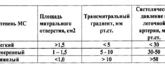
Fluid in the pleural cavity on objective examination. The ejection fraction has decreased to 40%. The end-diastolic size increased. The left atrium remained large. Pulmonary artery systolic pressure was high: 50 mmHg. pillar
Regurgitation was clearly detected on the mitral and tricuspid valves by auscultation, which was confirmed by echocardiographic examination to the 3rd degree.
Consequently, we had before us a patient with a clear clinical picture of both left and right heart failure. With swelling of the lower extremities. With low ejection fraction. With dilatation of almost all chambers of the left ventricle, right atrium, left atrium and high pulmonary hypertension.
06:18
(Slide show).
During this period, an electrocardiogram was obtained, which, of course, would have alarmed any doctor. You see (here especially in leads V1, V2, V3) electrical alternans. We see a blockade of the conduction pathways, overload of the left and right parts of the heart.
What attracts attention.
Look, in chest leads V4, V5 and V6 we see a pathological Q wave and ST segment elevation.
Such an electrocardiogram raised the question of the need for a differential diagnosis between myocardial infarction and those non-coronary myocardial diseases that can occur with such severity and severity, clinical and electrocardiographic. In particular, we could be talking about myocarditis.
During an echocardiographic study in May 2008, we see that there is no myocardial hypertrophy. Interventricular septum – 11 mm, posterior wall – 10 mm, end-diastolic size – 65. Ejection fraction – 39%. At the same time, accumulation of fluid in the pleural cavities was visible.
The same study - apical four-chamber position. We see large left and right atria. Simpson ejection fraction – up to 15%. These are very serious indicators against the background, as you already remember, of a clinical picture of congestive heart failure and serious changes in the electrocardiogram.
08:21
Thus, in addition to myocardial infarction, which I talked about, we were talking about a differential diagnosis:
• infectious myocarditis;
• hypersensitive myocarditis;
• giant cell myocarditis;
• cardiac lesions as part of a systemic disease (for example, polyarteritis nodosa);
• acute myocardial infarction.
Since we were talking about the possibility of hypersensitivity myocarditis, we carefully asked the patient what medications she had received before developing this disease.
It was a young patient, absolutely healthy. The only thing this patient took was contraception. Moreover, she took them for a long period: more than 10 years.
The last of the contraceptive drugs that “preceded” her illness was Novynette. A combination drug containing estrogen and estradiol.
The only thing that could be associated with, if we were talking about hypersensitive myocarditis, is with this contraceptive.
If we talk about different types of myocarditis. Assuming infectious myocarditis, we would have to rule out viral, bacterial, or septic etiologies. In autoimmune myocarditis, we would have to find antibodies to the myocardium.
Idiopathic giant cell myocarditis is usually observed in autoimmune diseases.
Acute hypersensitive myocarditis and acute necrotizing myocarditis are myocarditis that develop as a result of sensitization by previous use of certain drugs.
10:30
Monitoring of the patient (you see, a young patient of 35 years old) continues, starting in May.
The patient takes a relatively small amount of medications throughout this time. She was prescribed angiotensin-converting enzyme inhibitors "Diovan", "Carvedilol", "Verospiron", "Lasix" and "Metypred". ").
Of these drugs, she herself chose two for herself, which she took on an outpatient basis, increasing or decreasing their doses herself. It was very difficult to control the intake of these drugs.
She was taking Lasix. Against this background, she was able to reduce the duration and intensity of the cough. On the background of Metipred and Lasix, her shortness of breath decreased, weakness and pain in the right knee joint decreased. Swelling of the legs and feet decreased to a certain extent.
This, naturally, was one of the forms of diagnosis ex juvantibus - such a positive reaction (subjective and objective) to taking Metipred and a diuretic drug.
The temperature remained at low levels. It exceeded 37?C: 37.1?C, 37.2?C, 37.3?C. This continued until February and beyond - throughout 2009. Until October 2010, when she was admitted to our clinic again.
What attracts attention. As a child, the patient often suffered from sore throats. But these frequent outbreaks of tonsillitis, nevertheless, did not lead to the formation of valvular heart disease in the patient. In all likelihood, there was no talk of rheumatic fever.
She is a psychologist by profession. She went on vacation to Turkey several times. She smoked approximately 10 cigarettes a day for the past 5 years.
From family history. My grandmother died at the age of 35. Diagnosis unknown. My aunt has a congenital heart valve pathology. The patient's parents are healthy.
13:20
Rejection of the doctors’ recommendations forced us to organize a meeting between a psychiatrist and our patient in the very first days of her stay in the clinic. The psychiatrist, naturally, did not discover any mental illness, but still noted some character traits.
What are these features?
Anosognosia. The patient was not fully aware of her illness. She did not realize the severity and possible irreversible consequences of her condition.
The patient showed a distorted internal picture of the disease. The patient hoped for a miracle: something would happen that would bring her out of this unpleasant and difficult state.
Finally, low adherence to pharmacotherapy and non-pharmacological measures was identified. In particular, the patient, despite such a serious condition, continued to work. She did not follow the work and rest schedule.
Upon direct examination, a condition of moderate severity was noted. She is asthenic. You see, the mass is 55 kilograms. Height – 168. BMI – 19.5. The patient herself very scrupulously controlled her body weight and did not allow it, as she did.
Cervical lymph nodes that were not fused to the skin were palpated. Swelling of the legs and feet. The jugular veins swelled and pulsated to a level of 2 cm above the collarbone. Positive Plesh symptom.
Percussion above the lungs revealed a clear pulmonary sound up to the level of the 10th intercostal space, and then a dull sound that determined the fluid level. Bilateral hydrothorax.
Respiratory rate – 20. Mild tachypnea. In the lungs, vesicular breathing is harsh or weakened. In the basal regions, soft moist rales were heard on both sides.
Blood pressure is normal. The boundaries of relative cardiac dullness are somewhat shifted - this time to the left. The diameter of relative cardiac dullness is increased to 16 cm.
Rhythmic pulse. The first tone is weakened by auscultation. The protodiastolic gallop rhythm was again auscultated. On auscultation, there was a significant accent of the second tone over the pulmonary artery, which intensified with inspiration.
It must be said that during the examination of the patient, a paradoxical pulse was determined - a decrease in the amplitude of the pulse during inspiration. The liver protruded 5 centimeters from under the edge of the costal arch. Pasternatsky's symptom was negative.
16:40
There were no significant pathological changes in clinical and biochemical blood tests, with the exception, perhaps (we have already paid attention to this in retrospect), that in all studies the patient’s eosinophil level was maintained somewhere around 4%, 5%, 6% . Slight tendency to blood eosinophilia.
Since we differentiated between previous acute myocardial infarction and subacute current myocarditis (one of the forms of myocarditis), naturally, we conducted research.
The patient had very high activity and high levels of C-reactive protein. All other studies (anti-DNA antibodies, antinuclear antibodies, antineutrophil, cytoplasmic antibodies) were negative.
Antibodies to cardiolipin, which is very important, were also negative (IgM and IgG). This, in principle, could allow us to exclude autoimmune myocarditis in our patient. To a large extent, it probably allowed us to exclude giant cell myocarditis, because we were still unable to identify any signs of a rheumatological nature.
The only thing, if you pay attention, our patient has a slight increase in immunoglobulin E. This is evidence of hypersensitization of the immune system. In particular, we are talking about hypersensitization of cells of the innate immune system: macrophages, possibly producers of immunoglobulin E.
A very small combination, slight blood eosinophilia in combination with a slight increase in immunoglobulin E still gave us reason to assume an increase in sensitivity - about hypersensitivity that exists in our patient.
Diagnosis of pathology
At the initial appointment, the cardiologist examines patients, recording the presence of swelling of the neck, lower extremities, changes in the condition of the skin, pulse, and breathing processes. The collection of anamnesis is supported by the results of instrumental studies:
- electrocardiography;
- echography;
- X-rays of the lungs;
- magnetic resonance imaging.
Laboratory blood tests are also carried out, including biochemical, immunological, and bacterial culture. If necessary, the examination scheme may involve other diagnostic procedures, for example, histological analysis of myocardial tissue samples.
Why does myocarditis develop?
The majority of patients with myocarditis are young people under the age of 45. Women suffer more often, but their pathology occurs in a milder form due to the characteristics of the endocrine system. Since the chronic form of myocarditis is rarely accompanied by characteristic symptoms, patients often seek medical help in advanced stages, when the inflammatory process is complicated by serious arrhythmia, heart failure or vascular thrombosis. In the acute form, shortness of breath, severe swelling of the legs, and sudden attacks of arrhythmia are observed. Like most cardiac pathologies, this significantly increases the risk of premature death.
- In most cases, myocarditis is triggered by a viral or bacterial infection: tonsillitis, influenza, diphtheria, measles or others. Adenoviruses, streptococci, herpes, pathogenic fungi, salmonella, and trichinella can provoke an infection of the heart.
- Less commonly, myocarditis develops as a result of connective tissue diseases, autoimmune processes: systemic lupus erythematosus, rheumatoid arthritis, vasculitis.
- Ionizing radiation, strong toxins in alcohol, and medications can also provoke inflammation of the muscular lining of the heart.
- Allergic systemic reactions of the body are another group of causes of cardiac disease.
- Myocarditis, the causes of which cannot be determined, is called idiopathic.
Depending on the developmental characteristics, focal and diffuse forms of the disease are distinguished. Myocardial inflammation can have mild, moderate and severe severity, progress and recur.
Publications in the media
Almost any term in Latin with the ending “it” means inflammation: bronchitis is inflammation of the bronchi, tonsillitis is inflammation of the tonsils, etc. Myocarditis is inflammation of the muscle tissue of the heart. Similar to inflammation of any skeletal muscle after a sprain or hypothermia, which is manifested by pain, impaired muscle function, local swelling and redness, pain and decreased contractility also occur in the heart muscle. Due to inflammation, the current of impulses through the conduction system of the heart slows down - blockade and heart rhythm disturbances develop.
Myocarditis can occur with any acute or chronic indolent infectious disease. Most often, myocarditis develops due to viral infections. Non-infectious factors that cause myocarditis include some medications, including antibiotics, sulfonamides, etc., as well as vaccines and inoculations. In addition, myocarditis often accompanies systemic connective tissue diseases, for example, systemic lupus erythematosus, rheumatoid arthritis, and vasculitis. Among the causes of myocardial inflammation, a special place is given to rheumatism, in which myocarditis is one of the main and leading manifestations of the disease along with endocarditis and pericarditis - inflammation of the inner and outer lining of the heart, respectively.
There is also idiopathic (that is, without an obvious known cause) myocarditis, which is called Abramov-Fiedler. Moreover, in the classic course of the disease, examination and blood tests do not reveal the infection that provoked myocardial inflammation.
According to the course, acute, subacute and chronic myocarditis are distinguished. In a chronic course, depending on the frequency of relapses, cardiac function can significantly decrease and chronic heart failure develops.
Quite often, myocarditis is not accompanied by significant symptoms of the disease and is recognized only during an instrumental examination, sometimes performed for another reason. Complaints may be limited to general weakness, chest discomfort, palpitations, a slight increase in body temperature - nonspecific complaints, for which not everyone goes to the doctor, and almost no one comes to the cardiologist.
In typical, clinically pronounced cases, there are complaints of prolonged pain in the heart area not associated with physical activity, weakness, increased fatigue, shortness of breath and palpitations even at rest, and interruptions in cardiac function. Body temperature is often subfibrile: 37-37.90C.
Myocarditis begins against the background of an acute infectious disease or shortly after it. The malaise intensifies and the above complaints appear. Sometimes the pain in the heart is persistent and long-lasting. Body temperature is normal or slightly elevated. The severity of symptoms is determined by the prevalence of the inflammatory process in the myocardium - how much of the heart muscle is involved in inflammation and begins to work worse - to contract. Due to a decrease in myocardial tone, the size of the heart increases, the normal functioning of the valves is disrupted, and cardiac output decreases. Myocarditis can be complicated by the development of acute heart failure - a reason for emergency hospitalization and intensive care.
Myocarditis can occur in two clinical forms: infectious-toxic (heart lesions appear during a period of severe intoxication against the background of infection) and infectious-allergic (the infectious agent triggers an excessive immune response of the body and the body’s own immune cells begin to fight and destroy their own cells in different organs , including hearts, which in a number of ways are a little similar to bacteria and viruses. Therefore, the immune system cannot distinguish body cells from harmful microbes. That is why infectious-allergic myocarditis can occur and continue after the end of the infectious process that caused it.
Idiopathic myocarditis
Idiopathic myocarditis, in which there is no clearly established cause for the development of inflammation in the myocardium, is characterized by a more severe, sometimes fulminant course with the rapid development of a large “flabby” heart and heart failure. Without timely help, most often, such myocarditis is fatal or requires an urgent heart transplant, which in our country, unfortunately, is not always possible. In a large heart, blood flow slows down significantly, valve function is disrupted, and cardiac output decreases. The walls of the heart are stretched so much that even after successful treatment, changes in the myocardium can be irreversible. In idiopathic myocarditis, both rhythm disturbances and the formation of blood clots inside the heart due to its expansion and turbulent blood flow, which can enter the brain and lungs, leading to fatal thromboembolic complications - pulmonary infarctions, strokes, etc., are deadly in idiopathic myocarditis.
Myocarditis in children
Myocarditis in a child, as well as in an adult, occurs as a result of an infectious factor. In children, due to the failure of the immune response to a number of bacteria and viruses that adults are more “familiar” with, even a mild infection can lead to generalization - complications in other organs, most often the heart and kidneys. Children are characterized by the presence of two types of myocarditis:
1. Congenital - in this case, from the day of birth, the child exhibits lethargy, pallor, shortness of breath, changes in the ECG, low blood pressure, weak sucking reflex, etc. The child does not gain weight. Jaundice prolongs due to insufficient blood supply to the liver. The situation requires immediate treatment. Most often, this condition is associated with an intrapartum infection - then antibiotics are quite effective and the process is reversible. If congenital myocarditis is associated with impaired immunity and occurs as part of an autoimmune process, treatment is more complex, the prognosis is worse, and death is possible.
2. Purchased. This type of myocarditis, as in adults, is divided into acute, subacute and chronic, and is most often a consequence of ARVI. The child's appetite decreases, anxiety occurs during the day and sleep disturbances at night, episodes of cyanosis (blue face) and shortness of breath, increased body temperature. In this case, examination and observation by a cardiologist is mandatory.
Instrumental examination methods
In a blood test, the level of inflammation indicators increases - leukocytes, ESR, C-reactive protein, etc., but, unlike a banal infection, with myocarditis the level of enzymes in the blood increases - MB-CPK and troponin. These enzymes are normally found inside the muscle cells of the heart. During inflammation, which leads, among other things, to disruption of the integrity of cell membranes, enzymes enter the blood and their level is proportional to the number of damaged cells. This happens when the myocardium becomes inflamed or damaged during a heart attack.
The ECG records certain changes that are characteristic specifically of myocarditis, although often difficult to distinguish from ischemic ones. Almost always, with myocarditis, ECG reveals disturbances in heart rhythm and conduction.
Ultrasound of the heart - decreased myocardial contractility, expansion of the cavities of the heart, accumulation of fluid around the heart and other specific changes.
Forecast
With Abramov-Fiedler myocarditis, myocarditis against the background of sepsis (severe generalized infection and damage to almost all organs), the prognosis for life is unfavorable - mortality up to 50-60%.
With infectious myocarditis, with timely and adequate treatment, in most cases, myocarditis is asymptomatic and ends with complete recovery. Occasionally, changes in ECG or cardiac ultrasound data will persist for a long time. Most often, there are no signs of previous myocarditis 2-3 months after the end of the disease. Other forms of myocarditis with an acute and subacute course in at least 1/3 of cases result in complete recovery, and mortality in Russia reaches 5-10%.
Prevention
Prevention is the prevention and timely effective treatment of infectious diseases. If signs of infection appear, you should consult your doctor. Of course, a runny nose can be treated at home, but without a doctor and a basic examination you will not be able to distinguish between a “lingering” runny nose and the onset of an inflammatory process in the heart. It is also important to treat infectious diseases. Do not stop therapy prematurely, including antibiotics, even if nothing bothers you anymore.
If you have had myocarditis, you need to be examined and treated with greater care and attention for any, even “harmless” infection. If you are planning to undergo any surgical intervention, including tooth extraction, warts or skin papillomas, etc., be sure to undergo antibiotic prophylaxis under the supervision of a doctor.
Indications for hospitalization
In this matter everything is extremely simple. Suspicion of acute myocarditis or exacerbation of chronic myocarditis is an undeniable indication for hospitalization. Myocarditis is not treated at home, no matter how severe it is. Severe arrhythmias and acute heart failure can develop suddenly, and you will miss the moment to begin adequate treatment.
Treatment of myocarditis is carried out exclusively in a hospital under the dynamic supervision of a doctor. Antibiotics and anti-inflammatory drugs, including hormonal ones, are prescribed, the pulse rate is reduced and, if necessary, drugs are prescribed against disturbances in heart rhythm and conduction.
Cardiologist, Ph.D. Zaikina Alexandra
Diagnosis of myocarditis
For diagnostics
of myocardium, instrumental and laboratory methods are used, as well as myocardial biopsy.
Instrumental methods include:
- ECG - rhythm and conduction disturbances may be detected for the first time, sometimes infarction-like changes (especially in the acute phase of inflammation);
- ECHO CG - disturbances in systolic (due to the death of cardiomyocytes) and diastolic (due to swelling of the walls of the left ventricle) myocardial function;
- MRI contrast with gadolinium clearly reveals edema of the heart wall (as one of the stages of inflammation), but can be false negative;
- Myocardial scintigraphy can be informative in cases of suspected sarcoid myocarditis, but in other cases it is not very specific.
Laboratory methods include:
- A clinical blood test that can reveal an increase in ESR, an increase in the number of leukocytes, and in some forms, eosinophils;
- A study of the value of the so-called cardiac-specific enzymes (troponin T and CPK) reveals their increase, but a negative result does not exclude the diagnosis of myocarditis;
- Determination of serum antimyocardial antibodies.
- Routine serological viral testing is not recommended; it may only be useful in diagnosing diphtheria or borreliosis myocarditis
The gold standard for diagnosing myocarditis is endomyocardial myocardial biopsy (EMB), which is performed only in specialized hospitals and according to strict indications. Indications for referral are determined by the attending physician.
Nature and symptoms of the disease
With myocarditis, an increase in heart size, a persistent decrease in blood pressure, and insufficient blood circulation are observed. Patients suffer from shortness of breath, chronic loss of strength, quickly get tired, experience dizziness and physical weakness. Symptoms may be transient or persistent.
In most cases, the disease is accompanied by rhythmic disturbances: increased or decreased heart rate, tachycardia, bradycardia. The heart may pound strongly or seem to freeze in the chest. Some patients have dull pain in the chest area.
In acute myocarditis, a disease complicated by heart failure, the skin of the face turns pale and acquires a bluish tint. The neck veins swell, the temperature can rise for a long time to subfebrile. Interruptions in the functioning of the heart are accompanied by increased sweating, respiratory disorders, and vegetative signs: a feeling of crawling sensations, numbness in the arms or legs. Patients may suffer from pulmonary congestion. In severe cases, attacks of atrial fibrillation occur, which are life-threatening.
In chronic myocarditis, the symptoms are often blurred and mild. Periodically, patients experience attacks of bradycardia or rapid heart rate, dizziness, and weakness. They can get tired quickly during physical activity or fast walking.
Rheumatic myocarditis is often accompanied by joint symptoms: aches, impaired mobility, swelling of the joints, and increased body temperature. The malignant course of the pathology has a rapidly progressive nature, accompanied by expansion of the heart muscle and damage to the pericardium.
All cardiac patients have an individual clinical picture. Therefore, it is possible to make a diagnosis of “myocarditis” only after a comprehensive examination. With long-term inflammation, organic damage to the heart increases: myocardial cardiosclerosis is formed with a significant decrease in the functions of the organ.
Myocarditis is inflammation of the heart muscle. The disease is usually caused by a viral infection, but can be caused by a bacterial or fungal infection, alcohol abuse, certain autoimmune disorders, and medications. Weakening of the body's defenses and autoimmune disorders are important predisposing factors to the disease.
Myocarditis occurs at any age, with equal frequency in men and women. Most often it occurs under the guise of the infection that caused it and may go unnoticed. In severe forms, cardiac failure develops, which reduces blood delivery to organs and tissues. Blood clots may also form, leading to a stroke or heart attack.
For mild forms of myocarditis, no special treatment is required; the underlying disease is treated. In complicated forms, treatment is aimed at restoring normal heart function and correcting immune disorders. In severe forms of heart failure and blockage of blood vessels by blood clots, surgical intervention may be required.
Synonyms Russian
Inflammation of the heart muscle.
English synonyms
Myocarditis.
Symptoms
Symptoms of myocarditis are varied and may vary depending on the cause and severity of the disease. Mild forms may be asymptomatic or disguised as a viral infection. In severe cases, signs of heart failure come to the fore.
Heart symptoms include:
- chest pain, often radiating to the left arm;
- palpitations;
- rapid pulse;
- shortness of breath during exercise, and then at rest;
- swelling in the ankles and feet;
- fatigue.
General manifestations of infection:
- weakness;
- sweating;
- headache;
- pain in joints and muscles;
- fever;
- a sore throat;
- diarrhea.
General information about the disease
Inflammation of the heart muscle can occur against the background of any infection, most often viral. Cases of non-infectious myocarditis are also quite common and can be caused by certain medications, harmful chemicals or radiation. In many cases, the exact cause cannot be determined.
The most common causes of infectious myocarditis include:
- viral infections, among which the most important are Coxsackie enterovirus, influenza A and B, measles, rubella, infectious mononucleosis and hepatitis B;
- bacterial infections, primarily staphylococcal, streptococcal and diphtheritic;
- some parasites, primarily trypanosomes and toxoplasma;
- fungi (candida, pathogens of aspergillosis and histoplasmosis).
Non-infectious factors causing myocarditis:
- medications (antibiotics, sulfonamides);
- narcotic substances (cocaine);
- excess thyroid hormones;
- renal dysfunction;
- serums and vaccines;
- connective tissue diseases (nonspecific ulcerative colitis, scleroderma, rheumatoid arthritis, rheumatism, systemic vasculitis, Wegener's granulomatosis, systemic lupus erythematosus);
- injuries and burns;
- factors of unknown origin (idiopathic myocarditis Abramov-Fiedler).
Regardless of the cause of the disease, the changes that occur in the heart muscle are of a similar nature. As a result of inflammation, myocardial edema develops and its blood supply deteriorates. Individual cells can be destroyed with the release of substances into the blood, including protein fragments, which are perceived by the body's defense system as foreign due to their similarity to the proteins of some bacteria and viruses. This leads to activation of the immune system and the development of a reaction to its own tissues. The severity of these reactions largely determines the severity of the disease.
According to the course, acute and chronic myocarditis are distinguished. The course of the disease largely depends on its cause. Infectious myocarditis is most often acute or subacute, sometimes recurrent, and rarely becomes chronic. Non-infectious myocarditis usually turns into a chronic progressive form with the development of complications such as increasing heart failure, heart rhythm disturbances, blood clots, and heart attack.
Who is at risk?
- Suffering from systemic and autoimmune diseases.
- Patients with rheumatism.
- Having undergone heart surgery.
- Pregnant women.
Diagnostics
Diagnosis of myocarditis is difficult due to the similarity of the clinical picture with many other diseases. One of the warning signs may be the appearance of shortness of breath two weeks after a viral infection. The diagnosis of “myocarditis” is made based on the medical history, laboratory data and special research methods. If an infectious nature of myocarditis is suspected, a blood test should be performed for certain viruses, bacterial or fungal infections. The scope of laboratory tests is determined by the attending physician based on the specific manifestations of the infectious process. In most cases, the decisive factor in making a diagnosis is a myocardial biopsy.
Laboratory research
- Complete blood count (without leukocyte count and ESR) Red blood cells and hemoglobin may be reduced in severe forms of myocarditis.
- Leukocytes. An excessive number of leukocytes with a shift in the leukocyte formula to the left (the appearance of young cellular forms in the blood) is observed in bacterial myocarditis. With myocarditis caused by a viral infection, significant changes in the leukocyte formula, as a rule, do not occur. The number of eosinophils may increase, indicating an allergic reaction.
- Rheumatoid factor is an antibody detected in the blood of people who suffer from certain connective tissue diseases. In myocarditis, a positive rheumatoid factor indicates the presence of an autoimmune component of inflammation.
- Antistreptolysin O - test for the presence of antibodies to streptococcus in the blood. Prescribed for suspected myocarditis of streptococcal origin.
- Total protein and protein fractions in the blood may be reduced in myocarditis. A significant decrease in total protein levels can be observed in severe forms of the disease.
- Alanine aminotransferase (ALT), Aspartate aminotransferase (AST). ALT and AST are enzymes mainly found in the cells of the liver, pancreas, and heart muscle. Their elevated levels in the blood serum may indicate destruction of myocardial cells.
- Fibrinogen. Fibrinogen is one of the main components of the blood coagulation system. Increased fibrinogen levels are typically observed during active inflammation in response to tissue damage. Fibrinogen produces another protein, fibrin, which is involved in the formation of blood clots. In myocarditis, elevated fibrinogen levels may indicate a risk of thrombotic complications.
- Markers of myocardial damage: troponin and creatine kinase-MB may increase during the acute phase of myocarditis. An increase in their level in the absence of clinical myocardial infarction is of diagnostic significance. Troponin I is one of the proteins present in cardiac muscle. When damaged, troponin is released and its concentration in the blood increases. Increases in troponin levels in myocarditis can be observed in the acute phase of the disease. Creatine kinase-MB is an enzyme found in the heart muscle and is released when its cells are destroyed. In myocarditis, just like troponin, it may indicate myocardial damage.
- Antibodies to the myocardium. An allergic or autoimmune component of inflammation is present in the vast majority of cases of the disease, both infectious and non-infectious in origin. The appearance of antibodies to the heart muscle is one of the main indicators confirming the diagnosis.
- A blood test for antibodies to individual pathogens may be prescribed to confirm the infectious nature of the disease. For myocarditis, blood tests for:
- Flu
- Hepatitis B
- Candidiasis
- Streptococcal infection
Additional tests for suspected myocarditis include:
- Ultrasound of the heart (echocardiography). Allows you to evaluate the size and function of the heart. An echocardiogram can detect an enlarged heart, signs of developing heart failure, the manifestation of which on echocardiography is a decrease in ejection fraction. It is estimated as the percentage of blood volume entering the aorta during heart contraction and remaining in it. The lower the ejection fraction, the more severe the degree of cardiac dysfunction. An indicator below 35% indicates a high risk of developing arrhythmias. Echocardiography can also help rule out other diseases that have similar symptoms.
- An ECG allows you to record changes in heart rate, disturbances in cardiac conduction and rhythm, and suspect previous myocarditis. In severe forms of myocarditis, changes on the cardiogram may resemble those during myocardial infarction.
- Cardiac magnetic resonance imaging (MRI). Allows you to see the position and size of the heart, its shape and structure. MRI can show signs of myocardial inflammation.
- Myocardial biopsy. Using a special instrument, you can take fragments of the heart muscle for examination under a microscope. In this case, characteristic signs of inflammation and edema in muscle tissue are visible, as well as destroyed myocardial cells in severe cases of the disease. The method has high diagnostic accuracy and allows you to confirm or refute the diagnosis of myocarditis in cases where other research methods are insufficiently informative.
- Coronarocardiography. A method in which a radiopaque substance is injected into the vessels of the heart using special catheters. The method is important for excluding coronary heart disease, which in many cases should be differentiated from myocarditis.
Treatment
Treatment of myocarditis depends on the cause that caused it and the stage of the disease. In mild cases, restriction of physical activity is indicated; in more severe cases, bed rest is indicated. Salt restriction is recommended. Drug treatment may include:
- Antibiotics - prescribed when the bacterial nature of myocarditis is confirmed
- Steroid hormones - to suppress the autoimmune reaction and the allergic component
- Drugs that lower blood pressure and affect heart rate may be prescribed to reduce the workload on the heart.
- Diuretics to reduce swelling
In severe cases with the development of chronic heart failure, a pacemaker may be required or a heart transplant may be considered.
Prevention
There is no specific prevention of myocarditis. Basic measures to prevent myocarditis include:
- Scheduled preventive vaccinations
- Limiting contact with persons suffering from acute viral infection
- Timely treatment of viral diseases
- Maintaining personal hygiene rules
- A healthy lifestyle that eliminates the risk of HIV infection, avoiding drugs, limiting alcohol.
Recommended tests
- Erythrocyte sedimentation rate (ESR)
- Leukocyte formula
- General blood analysis
- General urine analysis with microscopy
- Total protein in urine
- Fibrinogen
- Serum albumin
- Total protein in whey
- Protein fractions in whey
- Serum creatinine
- Urea in serum
- Rheumatoid factor
- C-reactive protein, quantitative
- Total immunoglobulin A (IgA) in serum
- Total immunoglobulin G (IgG) in serum
- Antibodies to myocardium
- Troponin I (quantitative)
- Creatine kinase MB
- Influenza virus A/B (influenza viruses A/B), RNA [real-time PCR]
- HBV, DNA quantitative [real-time PCR]
- Candida albicans, IgG, titer
- Mumps Virus, IgG
- Varicella Zoster Virus, DNA [real-time PCR]
- Epstein Barr Virus, DNA [real-time PCR]
- HIV 1.2 Ag/Ab Combo (determination of antibodies to HIV types 1 and 2 and p24 antigen)
- Streptococcus pyogenes, DNA [real-time PCR]