Description
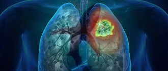
Pulmonary fibrosis is a process characterized by the replacement of lung tissue with fibrous (scar) tissue, which is accompanied by impaired respiratory function.
Thanks to the work of our lungs, the blood is saturated with oxygen necessary for energy consumption, as well as the release of carbon dioxide, which is formed as a by-product during the life of cells. The development of pulmonary fibrosis leads to a decrease in the volume of normally functioning lung tissue, and as a result, breathing efficiency decreases.
Replacement of pulmonary connective tissue can occur in one lung or in both at the same time. Depending on this, unilateral and bilateral fibrosis are distinguished. In addition, pulmonary fibrosis is divided into focal, in which a small area of lung tissue is affected, and total, in which the pathological process spreads to almost the entire lung.
The reasons for the development of pulmonary fibrosis are varied:
- diseases of the respiratory system (chronic bronchitis with broncho-obstructive syndrome, pneumonia, tuberculosis, chronic obstructive pulmonary disease);
- connective tissue diseases (rheumatoid arthritis, systemic lupus erythematosus, systemic scleroderma);
- exposure to production factors that negatively affect the respiratory system. For example, with prolonged inhalation of silicate dust in production, silicosis occurs. Occupational diseases also include asbestosis, which occurs due to inhalation of asbestos dust;
- long-term use of certain medications (antiarrhythmic drugs, drugs used to treat malignant tumors);
- the presence of vasculitis (a disease accompanied by inflammation of the walls of blood vessels);
- idiopathic or primary pulmonary fibrosis, the exact cause of which has not been established.
The prognosis of the disease is relatively unfavorable; progression of the pathological process leads to the development of pneumosclerosis and then cirrhosis of the lungs, which significantly reduces a person’s quality of life and can lead to disability. In addition, this condition leads to such serious complications as chronic respiratory failure, pulmonary hypertension, and chronic heart failure. Therefore, it is extremely important to contact a specialist as early as possible, who, in turn, will prescribe the necessary range of diagnostic procedures and necessary treatment.
General information
Fibrous changes in the lungs, what is it?
This is a pathological process of the formation of fibrous tissue (connective tissue) that replaces lung tissue, which leads to disruption of the elasticity of the lung and the function of gas exchange. With the formation of fibrous changes (synonymous with “pneumofibrosis”), elasticity is irreversibly lost. Fibrous changes in the lungs are the outcome of many diseases of the bronchopulmonary system, and also occur in other diseases. As pneumofibrosis progresses, a gradual restructuring of the lung tissue occurs - the structure of the acinus , sclerosis of the walls of the bronchioles develops, the capillary network becomes empty, and abnormal glandular structures are formed. These changes destroy and deform the lung tissue, and cyst-like expansions and fibrous fields are formed in this place. Areas of fibrosis can occur around the lymphatic vessels, in the interlobular septa, along the outflow of lymph from foci of former inflammation, in the tissue around the bronchi and blood vessels.
The further course of the disease and prognosis depend on the type of pulmonary fibrosis and the degree of its spread, since it determines the degree of impairment of respiratory function and gas exchange, as well as the severity of clinical manifestations. There are relatively favorable types of fibrotic changes and unfavorable ones. pulmonary hypertension quickly develop , which ultimately leads to a deterioration in the patient’s quality of life and a shortening of its duration.
Symptoms
Photo: physiatrics.ru
Initially, pulmonary fibrosis is manifested by general weakness, decreased ability to work, and increased fatigue. The skin becomes pale, in some cases a bluish tint can be noted. In particular, cyanosis is observed at the tips of the fingers and toes, in the area of the nasolabial triangle, and at the tip of the nose. There is also sleep disturbance, with increased sleepiness during the day and insomnia at night. Some patients complain of weight loss.
The main symptom that significantly reduces a person’s quality of life is shortness of breath. Shortness of breath is characterized by changes in the frequency and depth of breathing, in most cases accompanied by a feeling of lack of air. Initially, it appears during physical activity, then, as the process progresses, it begins to bother a person in a calm state. In addition, a cough may appear, mainly of a dry nature, but in some cases there is a wet cough with the release of a small amount of viscous sputum. The nature of the sputum is mucous, less often mucopurulent, which will indicate the addition of a secondary infection.
The long course of the process leads to a change in the shape of the fingers, which is called the “drumstick symptom”. The terminal phalanges of the fingers of the hands thicken, acquire a flask-shaped shape, and the nail plates take on the appearance of clock hands. When pressing on the finger, there is a feeling of mobility of the nail plate, which is explained by a change in the structure of the tissue between the nail and the underlying bone.
The large volume of damage to the lung tissue and the duration of the process lead to the development of heart failure. The so-called “pulmonary heart” is manifested by the following symptoms:
- swelling starting from the feet, then affecting the legs, thighs, etc.;
- progressive shortness of breath, occurring not only with minor physical activity, but also at rest;
- swelling and pulsation of the neck veins;
- periodic feeling of discomfort or pain in the heart area;
- sensation of palpitations or interruptions in heart function.
Forecast
The prognosis and life expectancy for pulmonary fibrosis depends on the disease against which it developed, the degree of progression of the fibrosis itself, its extent and the degree of involvement of the bronchial tree (meaning the formation of bronchiectasis, which aggravates the course of the disease). Among the prognostic criteria, the main one is the type of formation of pulmonary fibrosis.
Favorable types include fibrous changes in the form of cords in the central and peripheral interstitium of the lungs, as well as the atelectatic type of fibrosis. The latter is formed in places of organized pneumonia or at the site of long-term collapse of lung tissue as a result of closure of the bronchi. The occurrence of these types of pulmonary fibrosis does not affect perfusion and diffusion in the lungs and does not lead to the development of respiratory failure. Unfavorable types include changes in the form of acinar fibrosis and honeycomb lung, which always lead to impaired perfusion and severe respiratory failure. It is this factor that reduces the patient’s life expectancy.
Honeycomb lung and its significant prevalence are considered the most unfavorable prognostic sign. At the same time, importance is attached to the size of the “honeycombs” - large “honeycombs” or a mixed type of structure with alternating small and large cysts are considered more unfavorable. As for their localization, the lower zonal location is considered unfavorable.
The natural history of idiopathic fibrosis is associated with gradual changes in lung tissue and progressive fibrotic damage, which at the final stage takes on the appearance of a “honeycomb lung”. Increasing fibrosis progressively worsens the patient's condition and affects life expectancy.
It is difficult to answer the question of how long people live with this disease, since it depends on the state of pulmonary function. Terms can range from 2 to 6 years. Thus, in acute cases, patients live no more than 2 years, in subacute cases - from 2 to 4 years, and in chronic cases - from 4 to 6 years. The situation is aggravated by concomitant diseases that arise with age. Death is caused not only by the development of acute respiratory failure, but also by ischemic heart disease, pulmonary embolism or lung cancer .
With slowly progressing silicosis, the prognosis for life is positive, since the process of transition from one stage to another can last decades. There are cases when the progression of fibrosis is not detected at all - in such cases the prognosis for life is favorable. A severe complication of silicosis is spontaneous pneumothorax , but it is very rare in modern forms of silicosis. tuberculosis develops against the background of silicotic fibrosis , then the course of the disease is unfavorable and, in general, the prognosis depends on the severity of tuberculosis and silicosis, and the form of tuberculosis.
Diagnostics
Photo: medserviceisrael.ru
Diagnosis begins with collecting complaints and anamnesis of the disease. Attention is drawn to progressive shortness of breath, which occurs not only during physical activity, but also when at rest. When talking with the patient, it is important to carefully inquire about the presence of diseases that can lead to pulmonary fibrosis. Of particular interest are diseases of the respiratory system: pneumonia, tuberculosis, chronic obstructive pulmonary disease, chronic bronchitis complicated by broncho-obstructive syndrome, bronchial asthma. It should be taken into account that not only diseases of the respiratory system can lead to pulmonary fibrosis, therefore it is necessary to clarify the presence of systemic diseases (systemic lupus erythematosus, rheumatoid arthritis, systemic scleroderma), and the possibility of exposure to industrial factors.
After a conversation with the patient, the doctor begins an examination, during which attention is drawn to pale skin, the presence of cyanosis, and changes in the shape of the terminal phalanges of the fingers like drumsticks, if any. Then auscultation (listening) and percussion (tapping) of the lungs are performed. With a small amount of damage to the lung tissue, there are no specific changes during auscultation and percussion.
The next stage of diagnosis is to prescribe general laboratory tests (complete blood count, general urine test, biochemical blood test), which allow you to assess the general condition of the body. To determine the vital capacity of the lungs, the patient is sent for spirography. This is a unique method of assessing the condition of the lungs by measuring the volume and speed of exhaled air. This procedure is not difficult to perform, does not require prior special preparation from the patient, is absolutely painless and takes a few minutes. Spirography allows you to assess the functional reserve of the lungs.
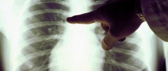
To visualize the pathological process, an X-ray examination of the chest organs is prescribed, which makes it possible to detect changes in the lungs. Changes are detected on both sides, mainly in the lower parts of the lungs. Strengthening and deformation of the pulmonary pattern are noted; a picture of the so-called “honeycomb lung” is gradually formed, which is characterized by the formation of ring-shaped shadows with a diameter of 3–7 mm with walls up to 3 mm thick, which to some extent resembles a honeycomb. Computed tomography (CT) and magnetic resonance imaging (MRI) provide a more detailed assessment of the structural condition of the lungs. In severe cases, when the above research methods do not allow making the correct diagnosis, a lung biopsy is used, which allows confirming the presence of scar tissue at the microscopic level. The material for research, namely a piece of lung tissue, is removed during an endoscopic examination or during surgery. This procedure is quite complicated to perform and can lead to various complications, so it is not currently used.
Classification
According to prevalence and X-ray or CT signs:
- Local pulmonary fibrosis (limited). Local pulmonary fibrosis affects only part of the lung tissue, which becomes denser. The pictures clearly define the boundaries of the pathological process. Limited pulmonary fibrosis does not affect the elasticity of the entire lung and has almost no effect on respiratory function. It is asymptomatic and often does not bother patients. An example of limited fibrosis would be the outcome of macrofocal pneumonia. In chronic bronchiolitis, X-ray examination reveals, in addition to local pneumosclerosis , thickenings around the bronchi. With this disease, bilateral fibrotic changes in the lower sections are usually found.
- Diffuse. With this option, fibrosis diffusely covers the entire lung tissue and the spread of the process occurs much faster than with the local form. The lung tissue becomes denser, the structure is deformed and the volume of the lungs decreases. In this regard, respiratory function deteriorates significantly.
- Focal pulmonary fibrosis is the presence of several foci of different sizes and structures. They can be either limited (rounded) or diffuse (blurred, without clear boundaries). Focal pneumofibrosis is detected in silicosis, pneumoconiosis, at the location of granulomas of various origins.
- Linear pulmonary fibrosis. In what pathology does it occur and what does “linear pulmonary fibrosis” mean? This term is used to describe an X-ray examination or CT scan of the lungs if a tissue compaction is detected in the form of a cord of a linear shape (practically it is a scar that has a linear shape). This type of fibrosis forms after inflammatory diseases (primarily tuberculosis ), injuries, and surgical interventions. Linear pathology often appears on x-ray only several years after the illness. An area of linear fibrosis, which is associated with the root of the lungs, indicates a history of hilar pneumonia or a primary tuberculosis complex. In the chronic form of exogenous alveolitis, CT manifestations are: cellular and rough linear fibrosis. In idiopathic fibrosis, parenchymal damage is symmetrical, and depending on the stage, linear fibrosis, ground glass, bronchiectasis and honeycomb lung are very often observed.
- Peribronchovascular fibrosis is the presence of connective tissue along the bronchovascular bundle. At the same time, the lumen of the bronchi and blood vessels narrows, which causes ventilation and vascular disorders.
- Acinar (intralobular) - the process reflects the filling of acini and terminal bronchioles with connective tissue cells. With this form, pronounced perfusion-diffusion disorders are noted. This is how interstitial pneumonia appears on CT.
- “Honeycomb lung” is a complete loss of the normal structure of the lung. In the lung tissue there is an alternation of fields of fibrosis, emphysema and cysts, which resembles a honeycomb.
With the flow:
- Progressive.
- Not progressive.
Idiopathic pulmonary fibrosis is a special form/variant of chronic fibrosing interstitial pneumonia of unknown etiology, which steadily progresses and is a cause of mortality. It occurs predominantly in older people and affects only the lungs. The process is more often localized in the peripheral parts of the lungs and is manifested by interstitial inflammation of the alveoli with the formation of fibrosis around the alveolar ducts.
As mentioned above, the causes of the disease are unknown, but there are factors that aggravate idiopathic pulmonary fibrosis - smoking, inhalation of inorganic and organic dust, viral infection, diabetes mellitus . The disease most often occurs in patients over 50 years of age. The incidence of the disease increases with age and predominates after 60–70 years.
Treatment
Photo: assets.copdnewstoday.com
Focal pulmonary fibrosis, which does not manifest itself clinically and does not cause discomfort to the patient, does not require treatment. Often, the focus of fibrosis is detected by chance during a preventive X-ray examination of the lungs. If it is detected, it is recommended to monitor the dynamics of the process and not delay contacting a specialist if symptoms from the respiratory system appear.
Total fibrosis requires the prescription of drugs with antifibrotic activity. The use of these drugs makes it possible to reduce the proliferation of fibrous tissue, which has a beneficial effect on the course of the disease.
In addition, we should not forget about the importance of specially designed physical training, which can improve lung function as much as possible, resulting in increased oxygen saturation in the blood.
Oxygen therapy (oxygen therapy) is used to eliminate oxygen deficiency. Oxygen is indispensable in the process of cellular respiration necessary for human life. With pulmonary fibrosis, blood oxygen saturation is significantly reduced. To compensate for this condition, courses of oxygen therapy are prescribed. However, it is also important to know that with frequent use of pure oxygen or inhalation mixtures with high oxygen concentrations, oxygen toxicity can develop. This condition is manifested by dry mouth, dry cough, chest pain, and in some cases there is a convulsive syndrome, which indicates hypertoxic damage to the brain. To prevent oxygen intoxication, it is important to strictly follow accepted standards and carefully monitor changes in the patient’s condition during an oxygen therapy session.
If the above treatment methods do not provide adequate effectiveness, the question of lung transplantation arises. There are certain indications for such a complex operation:
- significant decrease in vital capacity of the lungs;
- decrease in the diffusion capacity of the lungs by more than 2 times;
- respiratory failure in the stage of decompensation.
Such a solution to the problem in some situations is the only way to prolong a person’s life. According to statistics, most patients who have undergone lung transplantation have had their lives extended by 5 years or more. After such a complex operation, patients should periodically consult a therapist and pulmonologist. This is necessary to assess the general condition of the body and the functioning of the transplanted lungs.
List of sources
- Ivanova A. S. Fibrosing processes / A. S. Ivanova, E. A. Yuryeva, V. V. Dlin. M.: Overlay, 2008. 196 p.
- Idiopathic fibrosing alveolitis / M. M. Ilkovich [et al.] // In the book: Disseminated lung diseases / Ed. M. M. Ilkovich. M.: GEOTAR-Media, 2011. pp. 24–84. 3.
- Avdeev SN. Idiopathic pulmonary fibrosis: modern approaches to therapy. Practical Pulmonology 2015;1 22–31.
- Tsvetkova O.A., Voronkova O.O. Modern approach to the treatment of patients with idiopathic pulmonary fibrosis. Clinical Med 2017;95:281–5. 31.
- Aisanov Z.R., Kokosov A.N., Ovcharenko S.I., Khmelkova N.G., Tsoi A.N., Chuchalin A.G., Shmelev E.I. Chronic lung diseases. Federal program. RMJ, 2001; No. 1: p. 9 – 33.
Medicines
Photo: ravenna24ore.it
The main drugs used in the treatment of pulmonary fibrosis are:
- glucocorticoids;
- cytostatics;
- antifibrotic agents.
The administration of systemic glucocorticoids (prednisolone, dexamethasone) alleviates the general condition of the body. It is also believed that drugs in this group can influence the replacement of normal lung tissue with connective tissue, inhibiting this process. However, there is also a negative side to taking glucocorticoids. In the treatment of fibrosis, glucocorticoids are prescribed for a long period of time; in some cases, the course of treatment can reach up to 3 months. Such prolonged use is dangerous for the development of side effects. These include:
- increased blood pressure, which is especially dangerous for people suffering from arterial hypertension. While taking glucocorticoids, resistance to early antihypertensive therapy may occur;
- exacerbation of peptic ulcer of the stomach or duodenum;
- osteoporosis, which leads to increased bone fragility;
- weight gain;
- hyperglycemia. Therefore, when taking glucocorticoids, it is important to monitor glycemic levels. Particular attention should be paid to people with diabetes.
The appearance of these symptoms is the reason for contacting your doctor, who, in turn, either adjusts the dosage of the drug or discontinues it.
The use of cytostatics (azathioprine, cyclophosphamide) is also accompanied by some side effects: the function of the gonads is disrupted, hematopoiesis is inhibited, undesirable effects from the gastrointestinal tract, nephro- and hepatotoxicity are observed. The most gentle drug in this group in this regard is azathioprine. This drug is able to block cell division and degeneration of tissue into fibrous tissue, which has a significant role in the treatment of pulmonary fibrosis. Taking the drug is strictly prohibited during pregnancy and lactation, and is also undesirable if you have existing renal or liver failure.
Colchicine also belongs to antifibrotic drugs, which can inhibit the production of fibronectin. With long-term use, a picture of myelosuppression can be detected, that is, a decrease in leukocytes in the blood, and thrombocytopenia (decrease in platelets), temporary alopecia, myopathy, peripheral neuritis, etc. is not a rare case. The drug is contraindicated in cases of severe impairment of liver and kidney function, cardiovascular pathology, purulent infections, pregnancy and lactation. In all other cases, the use of the drug is justified.
Common reasons
- Inhalation of toxic substances.
- Exposure to radiation therapy , the consequence of which is post-radiation pulmonary fibrosis. Most often, post-radiation fibrosis develops after irradiation of the mediastinal area for lymphogranulomatosis and breast cancer . During radiation therapy, the development of pleural fibrosis is also possible. In the acute period after irradiation, pulmonary failure often develops, and in the later stages, radiation pneumonitis , intra-alveolar and interstitial fibrosis . Such complications are explained by the fact that the focus of irradiation falls on the area of the lungs, which are very sensitive to ionizing radiation and are quickly damaged. For radiation treatment of breast cancer, the dose is 70-80 Gy, and the maximum radiation tolerance of the lungs is 35-40 Gy. The incidence of complications depends on the total focal dose. In this case, the age of the patient and the location of the lesion are important. People over 70 years old have complications from radiotherapy 1.5 times more often than people aged 40-60 years. The closer to the mediastinum the focus is, the less often radiation damage to the lungs appears. Peripheral areas suffer more from radiation exposure.
- Taking medications: Methotrexate , Amiodarone , Proctolol (fibrosis can develop many months after the end of treatment with this drug), Propranolol (pleural fibrosis is possible). When taking nitrofurans, fibrosing alveolitis develops, and Methysergide and Ergotamine cause pleural sclerosis.
Folk remedies
Photo: narodnimisredstvami.ru
First of all, measures should be taken to prevent the development of pulmonary fibrosis. In many cases, pulmonary fibrosis is caused by diseases of the respiratory system, in particular pneumonia, which can lead to the replacement of lung tissue with connective tissue in the absence of treatment, the patient’s self-cancellation of antibacterial therapy, or failure to follow doctor’s recommendations. Therefore, it is extremely important, when the first symptoms of the disease appear, not to neglect your condition, but to consult a specialist and strictly follow his instructions. In addition, diseases such as chronic bronchitis with the phenomenon of bronchial obstruction, bronchiectasis of the lungs, chronic obstructive pulmonary disease, with a long course of the process, can also lead to the development of fibrosis. Considering this, it is important to avoid factors whose impact on the body leads to an exacerbation of the process. For the same purpose, annual influenza vaccination is recommended. People who are forced to use antiarrhythmic drugs for a long time need to be attentive to the appearance of symptoms from the respiratory system. If they are detected, you should immediately seek help from a doctor, and it is strongly recommended not to forget about an annual chest x-ray examination.
Special attention should be paid to people working in industries with occupational hazards. Long-term inhalation of organic and inorganic dust leads to the development of lung diseases, ultimately leading to fibrosis. Therefore, you should not neglect safety precautions, in particular the use of special devices that protect the upper parts of the respiratory system.
Traditional medicine recipes help to some extent alleviate the general condition of a person suffering from pulmonary fibrosis. For example, a decoction of rose hips and elecampane root has an excellent effect. To prepare it, grind the listed components, then pour 4 tablespoons of dry raw materials with 4 glasses of water and put on low heat. After boiling, keep it on the fire for another 15 minutes. Then the resulting broth is poured into a thermos and infused for 3 hours, after which it is carefully filtered. It is recommended to take half a glass 3 times a day before meals. The course of treatment is from 1 to 2 months. It is important not to interrupt the course of treatment and always have a ready-made remedy on hand. The combination of rosehip and elecampane helps to liquefy mucus, thereby making it easier to separate. In addition, this decoction protects lung tissue from inflammatory processes.
A recipe using anise seed is also widely used. To prepare this product, you should first prepare 1 tablespoon of anise seeds, which are poured with 1 glass of cold water. The resulting mixture is placed on the fire and brought to a boil, after which it is immediately removed from the stove. It is recommended to use the resulting product half a glass once a day.
The information is for reference only and is not a guide to action. Do not self-medicate. At the first symptoms of the disease, consult a doctor.
SEARCH FOR TREATMENT AROUND THE WORLD WITH YELLMED
Pathogenesis
The final sections of the respiratory tract consist of bronchioles (terminal and respiratory), acini and lobules. Terminal bronchioles are air-conducting, and respiratory bronchioles are involved in gas exchange, since they are connected to the alveoli through pores. The acini is the structure of the lung where gas exchange occurs and includes several respiratory bronchioles. The acini (from 3 to 24) are united into pulmonary lobules. The framework of the lobule includes interlobular septa, periarterial, peribronchial connective tissue, connective tissue of the alveolar and acini septa.
Connective tissue consists of collagen, reticular and elastin fibers. Interweavings of collagen and elastin fibers are present around the bronchi and in the alveolar walls, which determines the elastic properties of the lungs. Collagen provides rigidity to the intrapulmonary framework.
Connective tissue cells (smooth muscle and fibroblasts) synthesize elastin and collagen. Its primary role is to maintain the tone of the terminal sections of the respiratory tract. At the same time, connective tissue cells also synthesize enzymes (proteases) responsible for the destruction of elastin and collagen . The enzyme and antienzyme systems are in equilibrium under normal conditions. Under conditions of infectious inflammation, they continue to have an elastolytic (destructive) effect due to the high production of elastase (an enzyme that destroys elastic fibers, elastin, collagen). This causes the destruction of the normal connective tissue matrix, the destruction of the alveolar walls and the formation of common cavities. Excessive production of elastase or the absence of enzymes for its destruction is observed in a number of lung diseases ( COPD , emphysema , cystic fibrosis ).
In parallel, during inflammatory processes, there is an activation of fibroblasts that produce collagen. As a result, there is an enhanced process of development of connective tissue in the lung parenchyma and the formation of fibrosis. The development of fibrotic changes is preceded by edema and infiltration of the lung tissue. When the alveolar membrane is destroyed, intra-alveolar fibrosis develops at this site - growths of connective tissue spread into the alveoli. Then the connective tissue around the arteries and bronchi is involved in the pathological process.
Currently, a more acceptable theory of the pathogenesis of silicosis and other pneumoconioses is the immunological one, which states that the development of these occupational diseases is impossible without phagocytosis of dust particles by alveolar macrophages. The rate of their death depends on the fibrogenic aggressiveness of the dust and is proportional to it. The death of alveolar macrophages is a mandatory step in the formation of a silicotic nodule. The formation of a nodule is possible under the condition of repeated phagocytosis of dust. The resulting pro-inflammatory mediators ( cytokines ) provoke the accumulation of inflammatory cells in the septa of the alveoli, and oxygen radicals, damaging lung tissue, cause the death of macrophages.
Proteolytic enzymes (metalloproteinase and elastase) are released from destroyed macrophages and destroy lung tissue. The inflammatory phase is accompanied by recovery processes in which growth factors stimulate the formation of cells, epithelium and new vessels in damaged tissues. Uncontrolled epithelization processes lead to the development of fibrosis. Growth factors play one of the key roles in fibrogenesis . Their stimulating effect on the proliferation of fibroblasts and the destruction of collagen and fibronectin (high molecular weight protein of connective tissue) has been proven. In idiopathic fibrosis, fibrosis predominates with mild inflammatory changes. The main mechanism for the progression of pneumofibrosis is constant damage to the alveolar epithelium with impaired regeneration and excessive deposition of connective tissue due to the activation of fibroblasts.
Progression of fibrosis
The rate of progression of fibrosis varies markedly between patients. Among the known factors influencing the rate of development of fibrosis, the main ones can be identified - infection at an older age, male gender, alcohol abuse. At the same time, the connection between the viral load and the genotype of the virus and the rate of progression has not been established. The rate of fibrosis development is higher in immunocompromised patients. Fatty liver, obesity, and diabetes may also contribute to the faster development of fibrosis. To most accurately assess fibrosis progression, reassessment should be performed annually. In these cases, it is advisable to use methods for non-invasive assessment of liver fibrosis - informative and accessible ( FibroTest, FibroMax, elastomery ). Experience with the use of serum tests for assessing fibrosis and ultrasound techniques indicates the need for their combination for greater diagnostic accuracy.
Degrees of liver fibrosis
The severity of fibrosis in chronic liver disease reflects the long-term prognosis and, therefore, the need and urgency of treatment.
Liver fibrosis has 5 degrees (stages): F0, F1, F2, F3, F4 (cirrhosis). With viral hepatitis, for example, on average, it takes about 5 years from stage to stage. However, in later stages the rate of progression of fibrosis is higher. The rate of fibrosis development depends on the activity of the inflammatory process in the liver.
To determine the severity of fibrosis, there are various methods: biopsy, blood test, which measures biochemical markers of fibrosis formation ( FibroTest, FibroMax ). Currently, elastometry - direct ultrasound determination of the density of liver tissue using the Fibroscan device. The resulting density measurements at several points (10-20) in kiloPascals correspond to the degrees of fibrosis on the METAVIR scale from F0 - healthy liver, to F4 - cirrhosis.
A sufficient amount of scientific data has been obtained using this method and it has been proven that as the stage of fibrosis increases, the elasticity of the liver in kPa increases.
Legend:
- F 0-3 stages of fibrosis according to the METAVIR scale for chronic hepatitis,
- F 4 – cirrhosis of the liver,
- F 4+ URVP - cirrhosis of the liver with the presence of varicose veins of the esophagus,
- F 4+VRVP* - liver cirrhosis, portal hypertension, complicated by bleeding from varicose veins of the esophagus,
- HCC - hepatocellular carcinoma
Significance of differences p <0.05
Legend:
- F 0-3 stages of fibrosis according to the METAVIR scale for chronic hepatitis,
- F 4 – cirrhosis of the liver,
- F 4+ URVP - cirrhosis of the liver with the presence of varicose veins of the esophagus,
- F 4+VRVP* - liver cirrhosis, portal hypertension, complicated by bleeding from esophageal varices
The advantages of the method include simplicity, non-invasiveness, low cost, as well as clinical significance (prediction of complications of portal hypertension in liver cirrhosis, liver cancer formation). This determines broad indications for the method: initial examination, dynamics with and without treatment.
Limitations to the method are the presence of ascites, excess fatty tissue, and narrow intercostal spaces in the patient.
The results of Russian studies also confirmed high diagnostic accuracy (more than 80% for all groups) and reproducibility of the result by another operator.
The following elasticity thresholds are recommended for clinical use:
- 5.8 kPa to determine the boundary between stages F0 and F1 (METAVIR);
- 7.2 kPa to differentiate stages F1 and F2;
- 9.5 kPa – stages F2 and F3;
- 12.5 kPa to determine the boundary between severe fibrosis and cirrhosis of the liver - F4.
Factors influencing the diagnostic accuracy of elastometry:
- patient age >50 years,
- excess body weight,
- the presence of steatosis according to a morphological study of liver tissue.
Signs and symptoms of liver fibrosis
Early stage fibrosis is difficult to diagnose because it is often asymptomatic. Based on a blood test - the level of liver enzymes ALT and AST in the blood - one can judge the severity of fibrosis. AST levels are thought to have a stronger association with fibrosis than ALT levels. The AST/ALT ratio >1 is a reliable indicator of the severe stage of liver fibrosis (including liver cirrhosis).
The initial stage of liver damage due to fibrosis is characterized by an increase in liver size. Subsequently, the level of leukocytes, platelets and red blood cells decreases. As a result, the patient experiences anemia and thrombocytopenia. Signs that the disease is moving to the stage of cirrhosis are an enlarged spleen, varicose veins in the esophagus and hemorrhages from them.