The effectiveness of curiosin in trophic ulcers in patients with diabetes mellitus
P
It is generally accepted that in diabetes mellitus the wound healing process is significantly impaired, and the resulting trophic ulcers of various origins are difficult to treat. The existing different views on the pathogenesis of inhibition of the phases of the wound process do not reflect the true picture of the pathological process.
In clinical and experimental conditions, at the cellular level, we have discovered the mechanism of prolongation of the phases of inflammation, regeneration, scar formation and epithelization [3,4,5].
In diabetes mellitus, as a result of insulin deficiency, intracellular metabolic acidosis develops, an imbalance of macro- and microelements, the protein composition of the blood, hypercoagulation, and reduced cellular and humoral immunity. This condition is observed against the background of blood hypercoagulation and microangiopathy.
The shifts in homeostasis that we discovered distort the wound process, prolonging the time and phases of wound healing, both by primary and secondary intention, even with adequate insulin therapy (by 2 or more weeks).
If there is insufficient compensation for diabetes mellitus, these periods are postponed for a longer time, and ulcers of various origins can take weeks and months to heal.
We have found that in the first phase of the wound process - the stage of inflammation - the periods of resorption and rejection of necrotic tissue in the wound are prolonged, swelling and persistent neutrophilic infiltration of the walls and bottom of the wound or ulcer are observed for a long time (for 7-9 days).
In the second phase, the regeneration stage, the process of formation of argyrophilic and collagen fibers, inhibition of fibroblast proliferation and maturation of granulation tissue are delayed. In addition, the production of acidic mucopolysaccharides and hyaluronic acid decreases, the process of transformation of polyblasts and epithelial cells into fibroblasts slows down, and the synthesis of RNA and DNA decreases (10–12 days).
In the third phase of the wound process - the stage of scarring and epithelization, for a long time (10-12 days) the wound defect is filled with immature connective tissue, epithelization occurs slowly from the edges. A wide scar is formed, it is devoid of skin appendages and elastic fibers. [3]
These disorders are primarily associated with metabolic acidosis, because it is known that all enzymatic and oxidative processes in the cell, protein breakdown and synthesis, binding and release of oxygen are possible only if the pH of the environment is constant (about 7.4). This is also indicated by the detected ketone bodies in wound exudate [8].
It should be emphasized that the more severe the diabetes mellitus, the longer the phases of the wound process [5,6,7,9].
To speed up the healing process of wounds and trophic ulcers of soft tissues in patients with diabetes, antibiotics and enzymes, various water-soluble ointments, antiseptics, physiotherapeutic methods, etc. are used. [6,9]. However, the results of local treatment, even in a hospital setting, do not always satisfy both patients and doctors. The material costs are very high.
A special place in the treatment of trophic ulcers is occupied by organotherapeutic substances similar to the macromolecules of the body. These are substances of mucopolysaccharide nature that can interact with the cell membrane and hydrophobic proteins. Hyaluronic acid has this property. Combining it with zinc allowed the Gedeon Richter company to create the drug Curiosin, which we used for therapeutic purposes in patients with diabetes mellitus with long-term non-healing ulcers of various origins, in whom we clearly established a deficiency of hyaluronic acid in the cells of the body (wounds) [3, 5 ].
30 patients suffering from diabetes mellitus in combination with trophic, varicose and arterial ulcers of the lower extremities were examined. Three groups of 10 people were allocated.
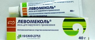
In the control group, a water-soluble ointment, levomekol, was used for treatment. For the remaining patients, a 0.2% solution of Curiosin from Gedeon-Richter was used, which was used in the form of dressings after cleansing the ulcers from necrotic masses.
According to the severity and duration of diabetes mellitus, age limit, gender, and duration of the ulcerative process, patients are distributed evenly in all three groups.
The study involved 19 men and 11 women aged from 65 to 81 years. Mild diabetes mellitus was noted in 7, moderate in 23. Duration ranged from 1.5 to 12 years. In practice, the ulcers existed from 4 months to 1.5 years. The area of the ulcerative surface ranged from 3.5 to 8 cm. The examination was carried out comprehensively, according to the program of the Gedeon-Richter company.
The state of red blood and leukocytes, glucose levels, measurement of the area of the ulcer, bacterial contamination and the composition of the microflora and cytograms of the wound process were performed according to generally accepted methods.
The only exception was the method for determining the number of microbial bodies in the wound. Based on the principle that in patients with diabetes mellitus, wounds and ulcers heal slowly and persistently, and gerontological patients refused to excise a piece of tissue from the bottom and edges of the ulcer, we applied a method for determining the number of bacteria not per 1 gram of tissue, but per 1 cm2 of the wound surface, according to the method developed at the Institute of Surgery named after. A.V. Vishnevsky [2].
In the process of a comprehensive examination of patients, data was obtained that allowed us to draw certain optimistic conclusions about the treatment of trophic ulcers.
Against the background of general corrective therapy, treatment of trophic ulcers with a 0.2% solution of Curiosin began 3–5 days after the bottom and edges were cleansed of necrotic masses. The bacterial composition of the surface of trophic ulcers did not vary widely. In 85% of cases, Staphylococcus aureus was sown, resistant to various broad-spectrum antibiotics (3–4 generation antibiotics). The number of bacterial bodies (b/t) per 1 cm2 in all 3 groups of patients indicated a high degree of infection of trophic ulcers that did not heal for months and often recurred.
The dynamics of bacterial contamination of trophic ulcers after 10 and 20 days of treatment are presented in Table 1.
In the control group of patients, in whom levomekol was used to treat trophic ulcers on the 10th day, the number of bacteria per 1 cm2 of surface ranged from 169 to 10,500 (on average 2400 ± 98.5 b/t).
In patients with varicose ulcers treated with curiosin solution, the average contamination was 2171 ± 171.2, and in patients with arterial ulcers (with contamination per 1 cm2 - 2443 ± 171.2), by 20 days the contamination of ulcers decreased by 10–12 times, which indicates a certain bactericidal effect of Curiosin.
Cytological prints of trophic ulcers in all three groups indicated the predominance of a purulent-necrotic process during the first 10 days of treatment. The first phase of the wound process (inflammation) was significantly longer compared to healthy people who did not suffer from diabetes. This was confirmed by our previous studies [3,4]. This was manifested in a decrease in wound discharge; the bottom and edges began to appear, first pale, and then juicy granulations. The bacterial flora decreased to 160–40 microbial bodies, the type of cytological picture was regenerative due to the appearance of connective tissue cells and a decrease in leukocytes. The inflammatory swelling of the tissues surrounding the ulcer decreased somewhat. When pressing on the skin, pain decreased, patients became calm, and psychological contact with others improved. By the 20th day of treatment, the marginal epithelization of the ulcer was clearly marked, the bottom was completely filled with succulent granulations, and it decreased in size. At the same time, in the control group of patients in whom levomekol was used, the healing (granulation and epithelization) of ulcers was prolonged.
In patients with diabetes mellitus who suffered from arterial ulcers of the lower extremities, the preparatory process for the administration of Curiosin was quite long (12–15 days). For this purpose, lysing enzymes (chymotrypsin), ultrasonic cavetation of a necrotic ulcer, and levomekol ointment were used. Sometimes it was necessary to perform necrotomy, amputation of fingers or fragments of the foot.
Old age, macro- and microngiopathies, neuropathies, diabetes mellitus and other concomitant diseases significantly changed tissue trophism and reduced the process of phagocytosis and granulation formation.
By the end of 10–12 days, when the first signs of inflammation and individual islands of pale granulations appeared, bandages with a 0.2% solution of Curiosin (1-2 drops per 1 cm2 of wound surface) were prescribed. The cytological picture was inflammatory and inflammatory-regenerative in nature for more than two weeks of drug use. At the same time, phagocytosis of the microflora was clearly expressed, and in a number of macrophages there was also an accumulation of individual cocci, with neutrophilic leukocytosis. Active proliferation of connective tissue cells was also noted.
In the control group of patients, where Curiosin was not used, the first and second phases of the wound process were 4-6 days longer compared to patients who received dressings with Curiosin.
With arterial trophic ulcers, the surrounding skin was pale, cold to the touch, atrophic, and there was no arterial pulsation in the feet. Often these patients used crutches or a wheelchair.
Upon admission to the hospital, most of our patients had severe swelling of the legs and feet, since due to severe pain they slept while sitting. After treatment, by day 20, swelling and pain decreased, patients became calmer, and began to sleep in a horizontal position.
Thus, complex corrective therapy (insulin, pentoxifylline, reapoliglucin, etc.) together with local application of Curiosin solution accelerates the process of cleansing and healing of trophic arterial ulcers by 6–7 days compared to the control group of patients.
The dynamics of changes in the area of trophic ulcers in the analyzed patients is also of interest (Fig. 1).
Rice. 1. Change in the area of the wound surface of trophic ulcers treated with Curiosin in patients with diabetes mellitus
From the presented graph it follows, firstly, that the initial area of the wound surface in both the control group and patients with ulcers of arterial origin was close in size: 49.4±3.2 mm and 42.5±2.7, respectively mm. In patients with varicose ulcers, in whom the disease lasts for years with frequent exacerbations, the average size of trophic ulcers was one third larger (62.7±5.3 mm); There was almost no concentric contraction of varicose ulcers due to the fibrous transformation of the subcutaneous tissue.
Secondly, arterial ulcers in combination with diabetes mellitus are very difficult to treat locally due to frequent microvascular thrombosis and cellular acidosis. Necrosis of the edges and bottom of the wound, often lined with tendon formations, requires a persistent and patient relationship between the doctor and the patient.
Thirdly, unlike varicose ulcers due to postthrombophlebic syndrome, arterial ulcers under the influence of a 0.2% Curiosin solution are capable of concentric contraction. Autodermoplasty in this case is ineffective, since the transplant becomes necrotic due to impaired peripheral blood flow. But, as noted above, trophic ulcers treated with Curiosin heal faster by a week or more compared to the control.
All of the above allows us to conclude that zinc hyaluronate (Curiosin) has a normalizing and accelerating effect on the formation of granulation tissue and epithelization of trophic ulcers in gerontological patients suffering from diabetes.
There is a deficiency of hyaluronic acid and the microelement zinc in the pancreas and other cells of the body [4,5]. This is one of the many reasons for the disruption of the regeneration process in diabetes mellitus. It should be noted that Curiosin has mild analgesic properties for varicose and arterial ulcers. The drug is very convenient to use both in the hospital and outside its walls.
The drug we used can be recommended for the treatment of trophic ulcers of various origins against the background of diabetes mellitus. For large ulcers with an area of more than 6–8 cm, Curiosin can be used with good effect to prepare the wound surface for autodermoplasty.
This is confirmed by data on healing by the 20th day of treatment with a 0.2% solution of Curiosin in 20% of ulcers of arterial origin, in 40% of venous ulcers and 10% in the control group of patients suffering from diabetes mellitus.
conclusions
1. Insulin deficiency, disrupting the trophic function of the body, significantly slows down the healing time of trophic ulcers of various origins.
2. Disturbances in the phase of the wound process are based on capillary circulation disorders (macro- and microangiopathies) and hypercoagulation, tissue metabolic acidosis, protein, electrolyte and microelement imbalances, reduced humoral and cellular immunity.
3. The use of a 0.2% solution of Curiosin, which is based on compounds of hyaluronic acid and zinc (and tissues and cells in patients with diabetes are poor in these components) is a good pathogenetic means of stimulating the healing of trophic varicose and arterial ulcers in this endocrinological pathology.
4. Actively influencing regeneration and epithelization, reducing treatment time by 10–12 days, 0.2% Curiosin solution can be effectively used to prepare the wound surface for autodermoplasty with large areas of the wound surface.
The list of references can be found on the website https://www.rmj.ru
Zinc hyaluronate –
Curiosin (trade name)
(Gedeon Richter)
References:
1. Gostishchev V.K., Kuleshov E.V., Mulyaev L.F. etc. Complicated osteoarthropathy is the main cause of lower limb amputations in patients with diabetes mellitus. In the book: wounds and wound infection. International conference, 1993; Part II, 333–5.
2. Kuzin M.I., Kolker I.I., Kostyuchenok B.M. Bacteriological diagnosis of wound infection. M., 1984; 22.
3. Kuleshov E.V., Shekhter A.B. Histochemical study of wound regeneration in diabetes mellitus. Archives of Pathology, 1972; 2:53–6.
4. Kuleshov E.V. Surgical diseases and diabetes mellitus. K. “Health”, 1990; 180.
5. Kuleshov E.V., Kuleshov S.E. Diabetes mellitus and surgical diseases. M., “Sunday”, 1996; 214.
6. Mc Murry JF Wound healing with diabetes mellitus. Surgeri Clin. North America, 1984; 64(4): 769–779.
7. Olefsky JU, Sherwin RS Diabetes mellitus. Management and complications. New York, 1985; 399.
8. Perile P., Holan J. The local exudative cellular response in ineoprolleit. Diabetes Clinical Research, 1961; 9 (2): 165–7.
9. Sanders C., Aldridge K. Current Antimicrobal Therapy of anaerobic infections. Europ J. Clin. Microbiol. Bis, 1992; 11(11): 999–1011


