Every spring and autumn I suffer from exacerbation of gastritis and pancreatitis, so at least once a year I go to the hospital, where they give me injections, give me pills and sometimes put on IVs. This disrupts the entire routine of life - problems at work (I’m the chief manager of a construction company, so I can’t be absent for a long time), in the family (the children are bored), and a lot of money is spent on so many medications. The decision to buy Dalargin came unexpectedly and out of nowhere. At the next appointment with the doctor, sitting and waiting for my turn, I met a woman who said that twice a year before the exacerbation season she undergoes a course of therapy with Dalargin and exacerbation does not occur. I asked a gastroenterologist about this medicine - she said that yes, indeed, you can use this advice.
When I came home, I found a description of Dalargin and was surprised that it was also effective for pancreatitis, but I had been to the doctor about chronic gastritis.
I bought the drug, the doctors gave me a prescription (dosage, course duration, how to administer it, etc.) and just at the end of February I started taking injections. I did the same thing in September and now do it all the time. To be honest, I forgot when I was in the hospital, definitely not for two years.
PS: The ampoules are not cheap, but compared to the amount that I usually shelled out for treatment at the clinic, this is mere trifle.
Dalargin - avoid fakes
Review left by Dgchuss
Rating: 3
(out of 5)
Advantages:
The real thing, and considering that the course of treatment is a maximum of a month, it’s also inexpensive
Flaws:
If you buy online, you might end up with a fake.
Sometimes I undergo therapy with Dalargin injections. I have a duodenal ulcer. The drug is administered intravenously. I usually inject myself at home, since my wife is a veterinarian, so she has experience in giving injections. Usually the reaction was normal and I always bought it online (cheaper than at the pharmacy). And then, after the first injection, my stomach began to growl loudly and diarrhea appeared. But the strangest thing is that it didn’t make me feel any better.
After that, I talked to the doctor and we came to the conclusion that most likely I had stumbled upon a counterfeit. I did not spare money and time searching for the site where I bought it before and ordered the required number of ampoules. This time there were no adverse reactions, but the effect was present. This is such a sad story, so look for the official website where the original Dalargin is sold.
UDC 616.3-089-06:616.37-002-08:615.243
The effectiveness of dalargin in the complex therapy of chronic pancreatitis that developed after gastric resection in patients with peptic ulcer was studied. A positive effect of dalargin on the clinical course of the disease and the functional state of the pancreas has been shown. The authors attribute this to the restoration of microcirculation and the antioxidant effect of the drug. According to long-term observations, the effect of therapy persisted for a year.
Evaluation of dalargin effectiveness in the treatment of postgastroresectional pancreatitis
The efficacy of dalargin in the treatment of chronic pancreatitis, which developed after stomach resection in patients with peptic ulcer disease, was investigated. The positive effect of dalargin on the clinical course of disease and the functional state of the pancreas was found. The authors attribute this to the restoration of microcirculation and antioxidant action of the drug. According to long-term observations the effect of therapy was maintained throughout the year.
Gastric resection remains one of the main surgical interventions for complicated gastric and duodenal ulcers [1]. However, after gastrectomy, 14.3-24.1% of patients develop postgastroresection pancreatitis (PP) [2, 3]. This may be a consequence of the shutdown of the antral hormonal zone and the duodenum, which play an important role in coordinating the activity of the digestive system [4, 5], as well as the result of a high load on the pancreas (P) due to its compensatory and adaptive function after gastrectomy. Prolonged pain syndrome and the development of exocrine pancreatic insufficiency significantly reduce the quality of life of patients and require serious drug therapy.
In recent years, natural peptides and their synthetic analogues have been recommended for the correction of various pathological processes, including in the pancreas. One of these regulatory peptides is the leu-enkephalin analogue dalargin [6, 7]. Clinical trials of dalargin conducted in hospital conditions demonstrated its high activity in the treatment of exacerbations of gastric and duodenal ulcers [8]. A positive therapeutic effect of the drug was noted in patients with acute pancreatitis [6, 7].
The purpose of our work is to study the characteristics of changes in the functional state of the pancreas after gastrectomy and to justify the use of dalargin in the treatment of patients with CP.
Materials and methods. We observed 50 patients with gastric and duodenal ulcers after gastric resection with subsequent development of PP, of which 27 patients underwent resection according to Billroth-1 and 23 according to Billroth-2. The study of patients was carried out from 1 to 31 years after surgery. The patients were aged from 26 to 71 years. There were 45 (90%) men, 5 (10%) women. The duration of peptic ulcer disease before surgery ranged from 1 to 39 years.
The patients were divided into 2 groups, comparable in age, severity, severity of pain, duration of pancreatitis, and frequency of concomitant pathologies: 25 patients in the observation group received dalargin daily in a dose of 1 mg intramuscularly 2 times a day as part of complex therapy, course dose of dalargin was 20-30 mg; 25 patients in the comparison group received traditional therapy, including antispasmodics, M-anticholinergics, proton pump inhibitors, and enzyme preparations.
The diagnosis was made on the basis of anamnestic, objective and laboratory data, ultrasonography, fibrogastroduodenoscopy, intragastric pH-metry and computed tomography. The exocrine function of the pancreas was assessed by the level of amylase, lipase and trypsin in the blood serum, and urine diastase; endocrine function - based on the level of insulin and C-peptide in the blood serum. Amylase in blood serum and diastase in urine were determined by the Caraway method, lipase was determined by a unified method. Trypsin, insulin and C-peptide, as well as TSH, T3, T4, cortisol and gastrin in the blood serum were determined by radioimmunoassay. The activity of lipid peroxidation processes was determined by the level of malondialdehyde (MDA) in blood serum. The state of microcirculation was assessed using conjunctival biomicroscopy at a slit lamp ShchL 2B BP according to the method of V.S. Volkova (1977).
The results of laboratory and instrumental studies of patients were compared with the data of 15 practically healthy individuals. The data were processed using the method of variation statistics; Student's t-test was used to assess intergroup differences; and the correlation analysis method was used to identify the relationship between indicators.
Results and its discussion
In the process of complex therapy with the use of dalargin, it was possible to completely relieve pain in 21 (84%) of 25 patients. A decrease in pain was observed mainly on the 3rd–4th day of therapy, and disappearance on the 5th–6th day. With traditional therapy, it was possible to completely relieve the pain syndrome in 72% of patients; the decrease in pain syndrome occurred mainly by the 5th–7th day of treatment.
Complex therapy with the use of dalargin helped to reduce or eliminate dyspeptic complaints. Nausea, a feeling of heaviness and fullness in the epigastric region disappeared most quickly in the observation group. The belching passed somewhat more slowly. An improvement in appetite was noted in 80% of the subjects in the observation group. Dalargin contributed to the normalization of stool in patients: constipation went away in all patients, mushy stool became formed. In the comparison group, nausea and bloating disappeared or decreased more slowly.
The use of dalargin clearly relieved pain on palpation of the abdominal wall in 84% of patients, in particular in the area of the projection of the pancreas, in the Shoffar, Gubergrits-Skulsky zone, but somewhat later than the reverse development of spontaneous pain syndrome.
In patients in the observation group, the basal level of blood amylase was elevated in 17.85% of cases, and decreased in 21.4% of cases. In the comparison group, an increased level of amylase in the blood serum was detected in 37.5% of cases, a decreased level - in 37.5%. In all patients with insufficient secretory function of the pancreas, ultrasound examination revealed an increase in the echogenicity of the organ structure, which indicates the development of sclerotic processes in the gland tissue as a result of previous exacerbations of the disease.
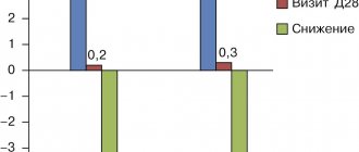
During the treatment, we observed multidirectional dynamics in the levels of blood enzymes (Fig. 1, 2).
Figure 1. Dynamics of amylase content with an initially increased level during therapy with dalargin (I) and traditional treatment (II)
Note: * - reliability in relation to the initial level;
**—reliability in relation to healthy people (p<0.05)
Figure 2. Dynamics of amylase with an initially reduced level during therapy with dalargin (I) and traditional treatment (II)
Note: * - reliability in relation to the initial level;
**—reliability in relation to healthy people (p<0.05)
In patients with a high level of amylase in the blood, its level significantly decreased during treatment in both groups; in patients with a reduced basal level of blood amylase, its increase was noted during a course of treatment with dalargin; the change in amylase level in the comparison group was insignificant (Fig. 2).
The increase in amylase levels can be explained by the stimulating effect of dalargin on pancreatic cells. Dalargin stimulates the processes of cell division and DNA synthesis, eliminates the development of erythrostasis and microthrombosis, improves microcirculation in the pancreas, and prevents the development of interacinar and intracellular edema in the intact segment of the gland [9-11].
A study of trypsin in blood serum revealed an increase in all examined patients with PP. During therapy, the concentration of trypsin in the blood in both groups decreased significantly (Table 1). Lipase activity in the examined patients was initially increased and decreased during treatment in both groups of patients.
Table 1.
Dynamics of lipase and trypsin levels in the blood of patients with PP during therapy
| Groups surveyed | Study period | Lipase (units) | Trypsin (ng/ml) |
| Comparison group | before treatment | 1,24±0,11* (n=11) | 620,2±9,8* (n=10) |
| after treatment | 0,64±0,09** (n=10) | 510,9±11,6** (n=11) | |
| Observation group | before treatment | 1,27±0,12* (n=9) | 840,6±9,8* (n=10) |
| after treatment | 0,5±0,07** (n=8) | 380,8±11,1** (n=9) | |
| Healthy | 0,78±0,14 (n=10) | 225,6±17,2 (n=10) | |
Note: n is the number of people examined;
* — reliability in relation to healthy people (p<0.05),
** — reliability in relation to the initial level
According to coprological studies, 23 patients (46%) had amilorrhea, 12 (24%) had creatorrhoea, and 7 (14%) had steatorrhea. During therapy, positive dynamics of the coprogram were noted: in the comparison group, the number of patients with creatorrhoea decreased by 50%, with amilorrhea - from 13 to 7 people, with steatorrhea - from 4 to 2 people; in the observation group, the number of patients with creatorrhoea decreased from 6 to 2 people, with amilorrhea from 10 to 4 people and with steatorrhea - from 3 to 1 person.
As the data in table shows. 2, in 55.8% of patients the insulin content in the blood was increased in comparison with healthy individuals, and in 33.2% of patients a decrease in its level was noted. A decrease in basal insulin levels was noted in patients with sclerotic changes in the pancreas identified by echography. Insulin secretion, both initially low and initially elevated, increased slightly in both groups of patients during treatment.
table 2.
Dynamics of insulin levels in the blood of patients with PN during therapy (mkd/ml)
| Group of examined people | Insulin level | Before treatment | After treatment |
| Comparison group | Initially reduced | 6,02±0,86 (n=8) | 6,92±0,41 (n=9) |
| Initially elevated | 16,25±1,25* (n=12) | 17,73±1,0 (n=10) | |
| Observation group | Initially reduced | 5,93±0,56* (n=7) | 6,46±0,41 (n=8) |
| Initially elevated | 12,88±1,06* (n=13) | 14,52±0,96 (n=11) | |
| Healthy | 8,83±0,96 (n=10) | ||
Note: n is the number of people examined;
* — reliability in relation to healthy people (p<0.05)
The ability of the pancreas to produce insulin is fully reflected by determining the concentration of C-peptide in the blood serum [12]. We did not identify significant differences in the level of C-peptide in the examined patients from healthy individuals: in the control group it was 0.3±0.09 pmol/ml, in the observation and comparison groups - 0.37±0.06 pmol/ml and 0 .34±0.03 pmol/ml, respectively. During therapy, a tendency towards an increase in the concentration of C-peptide in the blood serum was noted in both groups. The increase in insulin and C-peptide levels can be associated with the stimulating effect of therapy on islet tissue, improved metabolism of pancreatic tissue, and restoration of microcirculation. Thus, according to biomicroscopy of the conjunctiva, in the observation group the conjunctival index significantly decreased from 6.23 ± 0.68 to 4.21 ± 0.28 (in healthy people it was 1.88 ± 0.23) and in the comparison group it tended to decrease (from 6.26±0.66 to 5.33±0.44).
The gastrin level in the patients we examined was significantly higher in comparison with healthy individuals (Table 3). An increase in gastrin content in the blood may be due not only to atrophic lesions of the gastric stump mucosa, accompanied by a low concentration of hydrochloric acid, but also to a high content of trypsin in the blood, which promotes the formation of active gastrin [13]. The dependence of changes in gastrin levels on the type of therapy was established. Thus, in the comparison group there was a tendency towards an increase in its level, and during treatment with dalargin there was a tendency towards its decrease.
Table 3.
Dynamics of hormone levels in the blood of patients with post-gastroresection pancreatitis during therapy
| Group of examined people | Study period | Cortisol (nmol/l) | Gastrin (ng/l) | T3 (nmol/l) | T4 nmol/l) | TSH (med/l) |
| Comparison group | before treatment | 483,6±19,7* (n=12) | 66,86±7,5 (n=9) | 1,58±0,13 (n=11) | 118,2±9,8 (n=10) | 1,81±0,32 (n=8) |
| after treatment | 398.9±23.8 (n=9) | 71,95±4,9 * (n=10) | 1,81±0,17 (n=10) | 99,9±11,6 (n=11) | 1,7±0,29 (n=7) | |
| Observation group | before treatment | 480.3±21.7* (n=10) | 76,3±3,8* (n=7) | 1,8±0,09 (n=9) | 120,6±9,8 (n=10) | 2,31±0,41 (n=7) |
| after treatment | 461,2±18,0* (n=11) | 61,2±5,7 (n=13) | 2,01±0,2 (n=8) | 118,8±11,1 (n=9) | 2,1±0,34 (n=6) | |
| Healthy | 364,75±22,3 (n=15) | 52,8±3,91 (n=15) | 1,82 ±0,14 (n=10) | 124,68±17,2 (n=10) | 2,19±0,47 (n=10) | |
Note: n is the number of observations;
* — reliability in relation to healthy people (p<0.05)
According to our data, the basal level of cortisol in patients was higher than in healthy people (Table 3). During treatment, in both groups of patients there was a tendency towards a decrease in the level of cortisol in the blood. Considering that cortisol is one of the stress hormones [12, 14, 15], we are inclined to believe that hypercortisolemia in patients was largely of stress origin and decreased with relief of pain.
In patients, the basal level of T3 was lower, and no significant changes were noted in the content of T4 (Table 3). During treatment, an increase in T3 levels was observed in both groups of patients. The TSH level in patients with PP was practically no different from healthy individuals. The therapy in both groups of patients led to a decrease in its concentration in the blood.
The content of MDA in the blood, indicating the activity of lipid peroxidation processes, was increased in both groups of patients compared to healthy ones. After a course of therapy with dalargin, its amount in the blood decreased from 9.05±0.51 to 6.48±0.44 mmol/l (p<0.05), while in the group of patients receiving traditional therapy it changed immaterial. We attribute this to the pronounced antioxidant effect of dalargin.
To evaluate the effectiveness of therapy, according to long-term results, we monitored 31 patients for two years after the course of therapy, with 15 patients from the observation group and 16 patients from the comparison group. After hospital treatment, the patients were registered at the dispensary. Relapses of the disease occurred within two years in the comparison group in 9 (56.3%), and in patients in the observation group, exacerbation of the disease was noted in 6 (40%). At the same time, relapses of the disease in the comparison group more often occurred in the first months after discharge from the hospital and in general during the year their frequency was 43.8%. In the observation group, relapses of the disease occurred in the 2nd year after hospital treatment.
The majority of patients (39.5%) noted a violation of diet, physical activity and neuropsychic stress as the cause of relapse of the disease. In the comparison group of 9 patients with exacerbation of PP, 7 patients (77.8%) received inpatient treatment, while in the observation group 3 (50%) of 6 patients were inpatiently treated.
conclusions
1. When dalargin is included in complex therapy in patients with PP, pain and dyspeptic symptoms are eliminated or reduced, and at the same time a number of positive changes are noted in the functional state of the pancreas.
2. During therapy, the elevated levels of gastrin and cortisol in the peripheral blood decrease.
3. A pronounced antioxidant activity of dalargin was revealed, which may be one of the pathogenetic mechanisms of its therapeutic effect.
4. The use of dalargin for PP, according to short-term and long-term observations, was superior in effectiveness to traditional therapy.
ME AND. Grigus, O.D. Mikhailova, V.F. Bulychev, A.Yu. Gorbunov
Izhevsk State Medical Academy
Grigus Yan Ilyich - Candidate of Medical Sciences, assistant at the Department of Propaedeutics of Internal Diseases with a course in Nursing
Literature:
1. Zhizhin F.S., Kapustin B.B. Single-row antireflux anastomosis during tubular gastrectomy // Conference materials. "Current aspects of hospital surgery." - Izhevsk, 2000. - pp. 21-24.
2. Pomelov V.S., Baramidze G.G. Diagnosis, prevention and treatment of post-gastroresection reflux gastritis // Surgery. - 1994. - No. 5. - P. 32-35.
3. Vakhrushev Ya.M., Ivanov L.A. Postgastroresection syndromes. - Izhevsk, "Expertise", 1998. - 139 p.
4. Ugolev A.M. Enterin (intestinal hormonal) system. - Science, L., 1978, 315 p.
5. Vakhrushev Ya.M. New approaches to the study of functional connections of the gastrointestinal tract with the endocrine glands // Ter. archive. - 1985. - No. 9. - P. 98-102.
6. Georgadze A.K., Buzenkov S.V., Dzhakiya A.K. and others. Features of diagnosis and treatment of fatty pancreatic necrosis // Surgery. - 1991. - No. 4. - P. 11-14.
7. Penin V.A., Titov M.I., Titov V.N. and others. Mechanism of action and prospects for the use of a new domestic analogue of enkephalin - dalargin in the complex treatment of acute pancreatitis // Sat. scientific works "Acute pancreatitis". - Moscow, MMSI, 1986. - pp. 20-25.
8. Alekseenko S.A., Timoshin S.S. The influence of histamine and dalargin H2 receptor blockers on the reparative processes of the mucous membrane of the gastroduodenal system in patients with duodenal ulcer // Klin. honey. - 1996. - No. 9. - P. 52-54.
9. Titov M.I., Vinogradov V.A., Bespalov T.D. and others. Dalargin is a peptide drug with a cytoprotective effect // Bulletin. VKSC AMS USSR. - 1985. - No. 2 - P. 72-76.
10. Vinogradov V.A., Polonsky V.M. Dalargin is the most active synthetic analogue of endogenous opioids for the treatment of peptic ulcer disease (results of a five-year search) // Bulletin. VKSC AMS USSR. - 1986. - No. 2. - P. 62-63.
11. Ivanikov I.O., Vinogradov V.A. Results of using dalargin for pancreatitis // Mater. conf. VNOG. - Smolensk, 1991. - P. 224-226.
12. Vakhrushev Ya.M., Trusov V.V., Vinogradov N.A. Liver and hormones. - Izhevsk, 1992. - 112 p.
13. Trefflot MJ, Laugier R, Brethols A et al. Increased gastrin release in chronic calcifying pancreatitis and in chronic alcoholism // Horm. Metab. Res. - 1980. - No. 12. - R. 240.
14. Clinical endocrinology (Guide for doctors) / ed. N.T. Starkova. - M.: Medicine, 1991. - 512 p.
15. Mikhailova O.D. Endogenous intoxication in chronic pancreatitis // Ros. Journal of Gastroenterology, Hepatology and Coloproctology. - 2010. - No. 5. - P. 65.
Dalargin as an adjuvant drug
Review left by Kirill
Rating: 4
(out of 5)
Advantages:
Works quickly and efficiently
Flaws:
Price
I have problems with the trigeminal nerve, so I take many medications from different groups at the same time. Unfortunately, some of them have a very negative effect on the gastrointestinal tract, especially acidic Carniton. In this regard, the doctor recommended that I additionally inject myself with Dalargin solution, as it reduces acidity, neutralizes proteolysis and secretion in the pancreas. It also speeds up metabolic processes and generally has a positive effect on the body.
To reduce acidity, which was my most important problem, I was offered other alternative options in the form of tablets, which would be much cheaper. But in terms of effectiveness, no drug can compare with Dalargin.
Antisecretory drugs in the complex treatment of acute pancreatitis
Acute pancreatitis is an acute aseptic inflammation of the pancreatic tissue of the demarcation type, which is based on acute dystrophy, enzymatic autoaggression with necrobiosis of pancreatocytes and subsequent outcome in necrosis of the glandular tissue and surrounding structures with the addition of an endogenous secondary purulent infection or in sclerosis of the pancreas with atrophy of its glandular apparatus .
In a modern emergency surgery clinic, acute pancreatitis ranks third in the number of emergency hospitalized patients, second only to acute appendicitis and acute cholecystitis. Despite the constant improvement of treatment tactics and the introduction of new diagnostic and treatment technologies, over the last decade, mortality in acute pancreatitis ranges from 7-15%, and in destructive forms of pancreatitis reaches 40-80%.
To date, a colossal number of works have been published on the problem of acute pancreatitis. However, until now, most of the provisions regarding the etiology, pathogenesis, classification and treatment tactics for this pathology remain highly controversial. It is generally accepted that acute pancreatitis is a polyetiological disease. At the same time, American authors consider alcohol abuse to be its main cause, German authors consider cholelithiasis, Chinese and Vietnamese authors consider ascariasis. However, traditionally there are two groups of etiological factors for this disease. The first group includes factors that determine the disruption of the outflow of pancreatic secretions from the acini along the intralobular ducts into the main pancreatic duct and further into the duodenum, which leads to a sharp increase in pressure in the ductal system of the pancreas (hypertension-ductal factors). Intraductal hypertension occurs with spasm, inflammatory, cicatricial and neoplastic stenoses of the major duodenal papilla, including the sphincter of Oddi, and choledocholithiasis. Spasm of the sphincter of Oddi can be a consequence of both various neuro-reflex influences from the receptors of the hepatogastroduodenal zone, and direct irritation of the sympathetic and parasympathetic parts of the nervous system. Excitation of the vagus nerve causes hypersecretion of pancreatic juice, spasm of the sphincter of Oddi, and the occurrence of stasis and hypertension in the pancreatic duct system. It has been established that long-term consumption of alcohol in relatively large doses directly causes an increase in pressure in the small ducts of the pancreas. Etiological factors belonging to the second group lead to primary damage to acinar cells under conditions of normal intraductal pressure (primarily acinar factors). It is known that primary damage to pancreatic acinar cells can occur with local hemoperfusion disorders, allergic reactions, metabolic disorders, hormonal imbalance, toxic effects, infections, and trauma to the pancreas. The role of the nutritional etiological factor of acute pancreatitis can be reduced to the following. Foods rich in proteins and fats, alcohol cause pronounced secretion of pancreatic juice, rich in protein and poor in bicarbonates, which, if there is inadequate outflow, can cause the development of alimentary pancreatitis. Enhanced secretory activity of acinar cells with excessive food irritations or when eating protein-poor food is accompanied by irreversible intracellular damage to the acinar apparatus and the development of metabolic pancreatitis. It has been noted that excessive consumption of protein-rich foods can lead to sensitization of the body with protein metabolites, which leads to the development of allergic pancreatitis. Thus, the main etiological factors of acute pancreatitis can be called the following: cholelithiasis, pathology of the terminal part of the common bile duct and BDS, alcohol abuse , injuries (including operating rooms) of the pancreas, vascular diseases, metabolic disorders, infections, intoxications, autoallergic conditions. It has been experimentally shown and clinically confirmed that the most severe forms of acute pancreatitis develop with a combination of three etiological factors:
pancreatic hypersecretion;
acute intraductal hypertension;
intratubular activation of pancreatic enzymes.
The pathogenesis of acute pancreatitis currently also remains the subject of heated debate. It is believed that the development of acute pancreatitis is caused by a violation of the intracellular formation and transport of pancreatic enzymes, as well as intraacinar activation of proenzymes by hydrolases. The triggering mechanism for pathological reactions, which are the basis of inflammatory-necrotic damage to the pancreas, is the release of activated pancreatic enzymes from acinar cells, which are normally present in the form of inactive proenzymes. At the same time, today it is generally accepted that the processes of autolysis are primarily caused by the action of lipolytic enzymes. Activation of lipases occurs when the latter's proenzymes come into contact with bile acids and enterokinases. This situation occurs with hydraulic destruction of the acini due to intraductal hypertension, which is mainly a consequence of pancreatic hypersecretion and biliary-pancreatic or duodenopancreatic reflux with stenosis or insufficiency of the sphincter of Oddi and duodenal hypertension. It is assumed that alcohol not only has a direct toxic effect on pancreatocytes, but also causes the formation of protein microconglomerates that occlude small pancreatic ducts. Note that pancreatic lipase does not damage a healthy cell. The damage is caused by the action of phospholipase A, leading to the destruction of cell membranes, which makes it possible for lipase to penetrate the cell. When this mechanism is implemented, loci of fatty pancreatic necrobiosis with a perifocal demarcation shaft are formed. If the pathobiochemical process is limited to this, then fatty pancreatic necrosis is formed. If, due to excessive accumulation of fatty acids in the gland tissue, the pH reaches 3.4-4.3, intracellular trypsinogen is transformed into trypsin. In this case, trypsin activates lysosome proenzymes, as well as other proteinases that cause proteolysis of pancreatocytes. Activated elastase lyses the walls of blood vessels and interlobular connective tissue bridges, which contributes to the rapid spread of enzymatic autolysis in the pancreas and surrounding structures. Under the influence of trypsin, all pancreatic proenzymes (elastase, carboxypeptidases, chymotrypsin proenzyme), proenzymes of the kallikrein-kinin system, fibrinolytic enzymes and hemocoagulation profactors are activated, which ultimately leads to local and general pathobiochemical disorders with a possible outcome in the form of multiple organ failure syndrome . It is customary to distinguish a pre-infectious stage of the disease, in which aseptic inflammatory and necrotic foci are formed, and a phase of infectious complications - infected pancreatic necrosis, infected pancreatic necrosis with pancreatogenic abscess, retroperitoneal phlegmon.
Thus, one of the fundamental aspects of the complex of pathological reactions united by the concept of “acute pancreatitis” is intraductal hypertension in the pancreas. In this case, the main component of the increase in intraductal pressure is the secretion (in some situations - hypersecretion) of pancreatic juice.
The traditional classification of acute pancreatitis, accepted by practical surgeons, is a clinical and morphological classification that distinguishes acute edematous pancreatitis and destructive forms of pancreatitis - fatty pancreatic necrosis, hemorrhagic pancreatic necrosis, and also provides for the possible development of early and late complications. S. F. Bagnenko, A. D. Tolstoy, A. A. Kurygin (2004) distinguish the following clinical forms of acute pancreatitis, corresponding to the pathophysiological phase of its course:
Phase I is enzymatic and comprises the first five days of the disease. During this period, the formation of pancreatic necrosis of varying extent, the development of endotoxemia (the average duration of hyperfermentemia is 5 days), and in some patients - multiple organ failure and endotoxin shock. The maximum period for the formation of pancreatic necrosis is three days, after this period it does not progress further. However, with severe pancreatitis, the period of formation of pancreatic necrosis is much shorter (24-36 hours). It is advisable to distinguish two clinical forms: severe and non-severe acute pancreatitis.
- Severe acute pancreatitis. The incidence is 5%, mortality is 50-60%. The morphological substrate of severe acute pancreatitis is widespread pancreatic necrosis (large focal and total-subtotal), which corresponds to severe endotoxicosis.
- Mild acute pancreatitis. The incidence is 95%, mortality is 2-3%. Pancreatic necrosis in this form of acute pancreatitis either does not form (swelling of the pancreas) or is limited in nature and does not spread widely (focal pancreatic necrosis - up to 1.0 cm). Non-severe acute pancreatitis is accompanied by endotoxemia, the severity of which does not reach a severe degree.
Phase II is reactive (2nd week of the disease), characterized by the body’s reaction to the formed foci of necrosis (both in the pancreas and in the parapancreatic tissue). The clinical form of this phase is peripancreatic infiltrate.
Phase III – melting and sequestration (starts from the 3rd week of the disease, can last several months). Sequesters in the pancreas and retroperitoneal tissue begin to form from the 14th day from the onset of the disease. There are two possible options for the course of this phase:
- aseptic melting and sequestration – sterile pancreatic necrosis; characterized by the formation of postnecrotic cysts and fistulas;
- septic melting and sequestration - infected pancreatic necrosis and necrosis of parapancreatic tissue with further development of purulent complications. The clinical form of this phase of the disease is purulent-necrotic parapancreatitis and its own complications (purulent-necrotic leaks, abscesses of the retroperitoneal space and abdominal cavity, purulent omentobursitis, purulent peritonitis, arrosive and gastrointestinal bleeding, digestive fistulas, sepsis, etc.) .
It should be noted that not all authors share the point of view on the evolution of pathomorphological changes in acute pancreatitis and suggest the possibility of the occurrence of a primary destructive process (hemorrhagic pancreatic necrosis) without previous acute edematous pancreatitis and fatty pancreatic necrosis. This may be due to the fact that patients, due to a certain social background, are hospitalized already at the stage of hemorrhagic pancreatic necrosis or in the presence of purulent complications. However, most researchers support the opinion of the continuity of the morphological phases of acute pancreatitis. Thus, M. Schein (2004) calls pancreatitis “a disease of four weeks.” And this is quite understandable, both from the point of view of pathomorphology and from the pragmatic point of view of a practicing American surgeon. Indeed, the first two weeks are persistent complex conservative treatment, in the subsequent period - surgical interventions from minimally invasive (laparoscopy, transparietal punctures) to very aggressive (necrosequestrectomy, omentopancreatobursostomy, opening of pancreatogenic abscesses and phlegmon of the retroperitoneal space). Since in this section the author did not aim to continue the discussion about treatment tactics for acute pancreatitis (primarily about the indications, timing and scope of surgical treatment), the main attention is paid to the issue of conservative treatment of patients with this pathology. It should be noted that, according to a number of authors (A. D. Tolstoy, 2003, M. Schein, 2004), it is pathogenetically based complex conservative therapy for acute pancreatitis that is crucial for the outcome of the disease. This is especially true for acute edematous pancreatitis, since it prevents the transition of this form of pancreatitis to pancreatic necrosis. No less relevant is intensive conservative therapy for already formed foci of fatty or hemorrhagic destruction, which in this case prevents the spread of inflammatory-necrotic foci to previously intact tissue. In addition, taking into account the primary asepsis of the process in acute pancreatitis in the initial period of the disease, from the standpoint of common sense, it is advisable to take active therapeutic treatment aimed at stopping pathological processes in the pancreas itself, preventing and treating pancreatogenic toxemia syndrome, and preventing purulent-septic complications.
Currently, the fundamental principles of conservative treatment of acute pancreatitis are set out in all guidelines for emergency abdominal surgery. Let us remind the reader of them with some comments. So, for acute pancreatitis the following are indicated:
- Measures aimed at suppressing the exocrine function of the pancreas: A) “Cold, hunger and rest” (local hypothermia, strict diet, bed rest); B) Drug suppression of pancreatic secretion: cytostatics (5-fluorouracil, tegafur), inhibitors of gastric secretion (antisecretory drugs - H2 blockers, PPIs), opioid receptor agonists (dalargin), pancreatic ribonuclease, somatostatin and its synthetic analogues (octreotide).
- Antispasmodic therapy: myotropic antispasmodics (drotaverine, papaverine), anticholinergics (platiphylline, atropine), infusions of a glucose-novocaine mixture.
- Measures aimed at inactivating pancreatic enzymes circulating in the blood and inhibiting the cascade of reactions of the kallikrein-kinin system: protease inhibitors - aprotinin, ε-aminocaproic acid.
- Pain relief: non-steroidal anti-inflammatory drugs, opioid (with the exception of morphine, of course) analgesics, regional novocaine blockades.
- Correction of hypovolemic and water-electrolyte disorders, improvement of microcirculation, inhibition of free radical oxidation: infusions of crystalloids, colloids (hydroxystarch preparations, gelatins), perfluororganic emulsions, albumin, fresh frozen plasma, specific and nonspecific antioxidants.
- Detoxification therapy and methods of afferent detoxification: dextran infusions, forced diuresis, extracorporeal detoxification (hemo-, lympho- and enterosorption, plasmapheresis, ultrahemofiltration).
- Replenishment of energy costs (at least 3500 kcal/day): parenteral nutrition, balanced enteral tube nutrition.
- Correction of enteral insufficiency syndrome: prevention or relief of intestinal paresis, decompression of the small and large intestine, enteral lavage, use of enterosorbents, antihypoxants.
- Preventive prescription of antibacterial drugs: third generation cephalosporins, fluoroquinolones, metronidazole, in case of developed pancreatic necrosis - carbapenems (meropenem).
- Posyndromic therapy.
The works of various authors over the past five to ten years clearly show the evolution of treatment tactics in patients with acute pancreatitis from aggressive surgical to conservative expectant. The modern approach to the treatment of patients with acute pancreatitis dictates the need to select a specific therapy option, taking into account the stages of pancreatitis, taking into account the dynamics of laboratory parameters and instrumental research data - ultrasound, computed tomography, magnetic resonance imaging.
It should be noted that an indispensable condition for the treatment of patients with any clinical and morphological form of acute pancreatitis is compliance with the main condition - creating rest for the pancreas. This is achieved by suppressing the production of enzymes by pancreatocytes, as a result of which the release of enzymes that lyse proteins (trypsin, chymotrypsin, elastase) and phospholipid cell membranes (phospholipases, cholesterol esterase) is significantly reduced. Thus, the resting state of the pancreatocyte promotes regression of autolysis and prevents necrotic changes in tissue. In this regard, in the complex therapy of acute pancreatitis, the leading place is occupied by drugs that directly or indirectly inhibit the exocrine function of the pancreas. The maximum therapeutic effect is achieved by synergistically suppressing the synthesis of enzymes at the level of the pancreas, removing and inactivating enzymes already circulating in the blood.
Historically, the first class of compounds used for this purpose in acute pancreatitis were cytostatics - 5-fluorouracil, tegafur. The disadvantage of these drugs is the inhibition of leukopoiesis, impaired immunogenesis, and the occurrence of hypo- and dysproteinemia. The use of these drugs is justified in cases of verified pancreatic necrosis in order to maximally suppress the secretory function of the pancreas and thereby reduce the level of pancreatic enzymes in plasma. Previously, drugs of the protease inhibitor class were widely used to inhibit pancreatic secretion, but it has now been established that protease inhibitor drugs are active only in the blood. Protease inhibitors, as a rule, do not enter pancreatic tissue in sufficient concentrations and cannot effectively perform their function in relation to pancreatic juice enzymes. In addition, protease inhibitors have an autoimmunizing effect. To suppress the exocrine function of the pancreas, the use of opioid receptor agonists (dalargin), which selectively accumulate in pancreatocytes and inhibit the synthesis of pancreatic proenzymes, is justified. Pancreatic ribonuclease has a similar mechanism of action, destroying the messenger RNA of cells, thereby inhibiting protein synthesis by pancreatocytes. The drugs of choice for acute pancreatitis include a synthetic analogue of the hormone somatostatin, octreotide, which has a pronounced inhibitory effect on the exocrine function of the pancreas due to the activation of specific D-receptors of pancreatocytes. The main directions of its action are inhibition of basal and stimulated secretion of the pancreas, stomach, small intestine, regulation of the activity of the immune system, cytokine production, and cytoprotective effect. In addition, octreotide acts in the same way on the parietal and chief cells of the stomach, helping to reduce acid formation. The usual dosage regimen for octreotide is 300-600 mcg/day. with three times intravenous or subcutaneous administration.
A pathogenetically substantiated method of inhibiting pancreatic secretion is the use of drugs that reduce gastric secretion - antisecretory drugs. To understand the mechanism of action of antisecretory drugs in acute pancreatitis, we should briefly dwell on the regulation of pancreatic secretion. Regulation of the secretion of pancreatic juice is carried out by neurohumoral mechanisms, with the main importance given to humoral factors - gastrointestinal hormones (secretin, cholecystokinin-pancreozymin), activated with the participation of releasing peptides secreted in the mucous membrane of the duodenum. Secretin enhances the production of the liquid part of the juice, and cholecystokinin-pancreozymin stimulates the enzymatic activity of the pancreas. Insulin, gastrin, bombensin, bile salts, serotonin also enhance the secretory activity of the gland. The secretion of pancreatic juice is inhibited by: glucagon, calcitonin, somatostatin. The process of pancreatic secretion includes three phases. The cephalic (complex reflex) phase is caused primarily by reflex excitation of the vagus nerve. The gastric phase is associated with the effects of the vagus nerve and gastrin secreted by the antral glands when food enters the stomach. During the intestinal phase, when acidic chyme begins to enter the small intestine, the rate of pancreatic secretion becomes maximum, which is primarily associated with the release of secretin and cholecystokinin by the cells of the intestinal mucosa. Thus, there is a direct connection between the secretion of hydrochloric acid by the parietal cells of the stomach, a decrease in intraduodenal pH, the production of secretin from the duodenal mucosa and an increase in the secretion of pancreatic juice. That is why, to inhibit the secretion of pancreatic juice, reduce intraductal pressure in the pancreas and, ultimately, to reduce intrapancreatic activation of enzymes, measures are used to suppress the secretion of hydrochloric acid in the stomach - a physiological stimulator of pancreatic secretion. A decrease in the acidity of gastric juice causes less pronounced acidification of the duodenum, as a result of which the secretion of secretin, the main hormone that stimulates the excretory function of the pancreas, decreases.
It should be noted that, despite the widespread (and in some clinics, obligatory) use of antisecretory drugs for the treatment of patients with acute pancreatitis, systematic studies on this issue have not been conducted either in Russia or abroad. From individual reports it is known that:
- the use of omeprazole in the complex treatment of patients with acute pancreatitis and exacerbation of chronic pancreatitis contributes to faster relief of abdominal pain syndrome, normalization of the clinical picture, relevant instrumental and laboratory indicators (Zvyagintseva T. D. et al., 2003; Minushkin O. N. et al. ., 2004) ;
— the clinical effectiveness of omeprazole in acute pancreatitis is the highest among antiulcer drugs. Omeprazole is highly lipophilic and easily penetrates into the parietal cells of the gastric mucosa, where it accumulates and is activated at an acidic pH. Rabeprazole has a shorter duration of action than omeprazole. For acute pancreatitis, the daily dose of omeprazole was 40 mg (M. Buchler et al., 2000);
- today, based on the principles of evidence-based medicine, we can confidently say that the effectiveness of PPIs in acute pancreatitis is significantly higher compared to H2-histamine receptor blockers (K. Bardhan et al., 2001, meta-analysis data by N. Chiba et al. ., 1999).
Considering the fact that to exclude acidification of the duodenum, the intragastric pH should not be lower than 4, the optimal regimen for using the parenteral form of omeprazole (Losec) for acute pancreatitis should be considered a bolus administration of 80 mg of the drug, followed by continuous infusion at a rate of 4 mg/h.
The need to use antisecretory drugs in acute pancreatitis is due to two more circumstances. Very often (in at least 20% of cases) acute pancreatitis is combined with peptic ulcer disease. In this case, the presence of at least one cause-and-effect relationship is obvious: ulceration - acute pancreatitis. Firstly, the development of an inflammatory-necrotic process in the pancreas is possible due to the penetration of ulcers into the head and body of the gland. Secondly, peptic ulcer disease, as a rule, is combined with severe disturbances in duodenal motility, which, realized through duodenal hypertension, leads to the formation of duodenal-pancreatic reflux. In these difficult clinical situations, control of gastric acid production is one of the main goals of treatment. Therefore, in this case, the use of antisecretory drugs, including long-term ones, has absolute indications. Finally, another indication for the prescription of antisecretory drugs in acute pancreatitis is the prevention of stress erosive and ulcerative damage, the need for which is especially relevant in severe acute pancreatitis with the development of large-focal pancreatic necrosis, purulent-septic complications and multiple organ failure syndrome.
In conclusion, we would like to emphasize once again that the use of a complex of modern intensive care measures (antisecretory therapy, other inhibitors of pancreatic secretion and proteolytic enzymes, detoxification agents) in patients with acute pancreatitis, taking into account the stages and individual dynamics of the disease, as well as timely prevention of purulent complications pancreatic necrosis will undoubtedly improve the results of treatment of patients with acute pancreatitis, shorten the hospital stay of patients, reduce the need for invasive treatment methods and, most importantly, reduce mortality.
Unusual use of Dalargin
Review left by Fatina
Rating: 5
(out of 5)
Advantages:
The versatility of the drug, the speed of manifestation of the effect
Flaws:
I didn’t find any special ones
I had granulosa pharyngitis, so I was very surprised when my attending physician at the hospital suggested taking a course of Dalargin injections. I know this drug firsthand, as my husband has a stomach ulcer and we buy ampoules from time to time. And I know for sure that it is for pancreatitis and ulcers. But I trusted the doctor, because she has a good reputation, and she has been working for more than 30 years.
So they injected me into the back wall of my throat, but somehow it was submucosal. I was surprised by the result - when I have pharyngitis I have a very, very strong cough, and one injection instantly neutralized it. After a course of 5 days, the cough disappeared. I didn't expect such an amazing effect. I give it 5+
Evaluation of the effectiveness of dalargin in the treatment of post-gastroresection pancreatitis
UDC 616.3-089-06:616.37-002-08:615.243
The effectiveness of dalargin in the complex therapy of chronic pancreatitis that developed after gastric resection in patients with peptic ulcer was studied. A positive effect of dalargin on the clinical course of the disease and the functional state of the pancreas has been shown. The authors attribute this to the restoration of microcirculation and the antioxidant effect of the drug. According to long-term observations, the effect of therapy persisted for a year.
Evaluation of dalargin effectiveness in the treatment of postgastroresectional pancreatitis
The efficacy of dalargin in the treatment of chronic pancreatitis, which developed after stomach resection in patients with peptic ulcer disease, was investigated. The positive effect of dalargin on the clinical course of disease and the functional state of the pancreas was found. The authors attribute this to the restoration of microcirculation and antioxidant action of the drug. According to long-term observations the effect of therapy was maintained throughout the year.
Gastric resection remains one of the main surgical interventions for complicated gastric and duodenal ulcers [1]. However, after gastrectomy, 14.3-24.1% of patients develop postgastroresection pancreatitis (PP) [2, 3]. This may be a consequence of the shutdown of the antral hormonal zone and the duodenum, which play an important role in coordinating the activity of the digestive system [4, 5], as well as the result of a high load on the pancreas (P) due to its compensatory and adaptive function after gastrectomy. Prolonged pain syndrome and the development of exocrine pancreatic insufficiency significantly reduce the quality of life of patients and require serious drug therapy.
In recent years, natural peptides and their synthetic analogues have been recommended for the correction of various pathological processes, including in the pancreas. One of these regulatory peptides is the leu-enkephalin analogue dalargin [6, 7]. Clinical trials of dalargin conducted in hospital conditions demonstrated its high activity in the treatment of exacerbations of gastric and duodenal ulcers [8]. A positive therapeutic effect of the drug was noted in patients with acute pancreatitis [6, 7].
The purpose of our work is to study the characteristics of changes in the functional state of the pancreas after gastrectomy and to justify the use of dalargin in the treatment of patients with CP.
Materials and methods. We observed 50 patients with gastric and duodenal ulcers after gastric resection with subsequent development of PP, of which 27 patients underwent resection according to Billroth-1 and 23 according to Billroth-2. The study of patients was carried out from 1 to 31 years after surgery. The patients were aged from 26 to 71 years. There were 45 (90%) men, 5 (10%) women. The duration of peptic ulcer disease before surgery ranged from 1 to 39 years.
The patients were divided into 2 groups, comparable in age, severity, severity of pain, duration of pancreatitis, and frequency of concomitant pathologies: 25 patients in the observation group received dalargin daily in a dose of 1 mg intramuscularly 2 times a day as part of complex therapy, course dose of dalargin was 20-30 mg; 25 patients in the comparison group received traditional therapy, including antispasmodics, M-anticholinergics, proton pump inhibitors, and enzyme preparations.
The diagnosis was made on the basis of anamnestic, objective and laboratory data, ultrasonography, fibrogastroduodenoscopy, intragastric pH-metry and computed tomography. The exocrine function of the pancreas was assessed by the level of amylase, lipase and trypsin in the blood serum, and urine diastase; endocrine function - based on the level of insulin and C-peptide in the blood serum. Amylase in blood serum and diastase in urine were determined by the Caraway method, lipase was determined by a unified method. Trypsin, insulin and C-peptide, as well as TSH, T3, T4, cortisol and gastrin in the blood serum were determined by radioimmunoassay. The activity of lipid peroxidation processes was determined by the level of malondialdehyde (MDA) in blood serum. The state of microcirculation was assessed using conjunctival biomicroscopy at a slit lamp ShchL 2B BP according to the method of V.S. Volkova (1977).
The results of laboratory and instrumental studies of patients were compared with the data of 15 practically healthy individuals. The data were processed using the method of variation statistics; Student's t-test was used to assess intergroup differences; and the correlation analysis method was used to identify the relationship between indicators.
Results and its discussion
In the process of complex therapy with the use of dalargin, it was possible to completely relieve pain in 21 (84%) of 25 patients. A decrease in pain was observed mainly on the 3rd–4th day of therapy, and disappearance on the 5th–6th day. With traditional therapy, it was possible to completely relieve the pain syndrome in 72% of patients; the decrease in pain syndrome occurred mainly by the 5th–7th day of treatment.
Complex therapy with the use of dalargin helped to reduce or eliminate dyspeptic complaints. Nausea, a feeling of heaviness and fullness in the epigastric region disappeared most quickly in the observation group. The belching passed somewhat more slowly. An improvement in appetite was noted in 80% of the subjects in the observation group. Dalargin contributed to the normalization of stool in patients: constipation went away in all patients, mushy stool became formed. In the comparison group, nausea and bloating disappeared or decreased more slowly.
The use of dalargin clearly relieved pain on palpation of the abdominal wall in 84% of patients, in particular in the area of the projection of the pancreas, in the Shoffar, Gubergrits-Skulsky zone, but somewhat later than the reverse development of spontaneous pain syndrome.
In patients in the observation group, the basal level of blood amylase was elevated in 17.85% of cases, and decreased in 21.4% of cases. In the comparison group, an increased level of amylase in the blood serum was detected in 37.5% of cases, a decreased level - in 37.5%. In all patients with insufficient secretory function of the pancreas, ultrasound examination revealed an increase in the echogenicity of the organ structure, which indicates the development of sclerotic processes in the gland tissue as a result of previous exacerbations of the disease.
During the treatment, we observed multidirectional dynamics in the levels of blood enzymes (Fig. 1, 2).
Figure 1. Dynamics of amylase content with an initially increased level during therapy with dalargin (I) and traditional treatment (II)
Note: * - reliability in relation to the initial level;
**—reliability in relation to healthy people (p<0.05)
Figure 2. Dynamics of amylase with an initially reduced level during therapy with dalargin (I) and traditional treatment (II)
Note: * - reliability in relation to the initial level;
**—reliability in relation to healthy people (p<0.05)
In patients with a high level of amylase in the blood, its level significantly decreased during treatment in both groups; in patients with a reduced basal level of blood amylase, its increase was noted during a course of treatment with dalargin; the change in amylase level in the comparison group was insignificant (Fig. 2).
The increase in amylase levels can be explained by the stimulating effect of dalargin on pancreatic cells. Dalargin stimulates the processes of cell division and DNA synthesis, eliminates the development of erythrostasis and microthrombosis, improves microcirculation in the pancreas, and prevents the development of interacinar and intracellular edema in the intact segment of the gland [9-11].
A study of trypsin in blood serum revealed an increase in all examined patients with PP. During therapy, the concentration of trypsin in the blood in both groups decreased significantly (Table 1). Lipase activity in the examined patients was initially increased and decreased during treatment in both groups of patients.
Table 1.
Dynamics of lipase and trypsin levels in the blood of patients with PP during therapy
| Groups surveyed | Study period | Lipase (units) | Trypsin (ng/ml) |
| Comparison group | before treatment | 1,24±0,11* (n=11) | 620,2±9,8* (n=10) |
| after treatment | 0,64±0,09** (n=10) | 510,9±11,6** (n=11) | |
| Observation group | before treatment | 1,27±0,12* (n=9) | 840,6±9,8* (n=10) |
| after treatment | 0,5±0,07** (n=8) | 380,8±11,1** (n=9) | |
| Healthy | 0,78±0,14 (n=10) | 225,6±17,2 (n=10) | |
Note: n is the number of people examined;
* — reliability in relation to healthy people (p<0.05),
** — reliability in relation to the initial level
According to coprological studies, 23 patients (46%) had amilorrhea, 12 (24%) had creatorrhoea, and 7 (14%) had steatorrhea. During therapy, positive dynamics of the coprogram were noted: in the comparison group, the number of patients with creatorrhoea decreased by 50%, with amilorrhea - from 13 to 7 people, with steatorrhea - from 4 to 2 people; in the observation group, the number of patients with creatorrhoea decreased from 6 to 2 people, with amilorrhea from 10 to 4 people and with steatorrhea - from 3 to 1 person.
As the data in table shows. 2, in 55.8% of patients the insulin content in the blood was increased in comparison with healthy individuals, and in 33.2% of patients a decrease in its level was noted. A decrease in basal insulin levels was noted in patients with sclerotic changes in the pancreas identified by echography. Insulin secretion, both initially low and initially elevated, increased slightly in both groups of patients during treatment.
table 2.
Dynamics of insulin levels in the blood of patients with PN during therapy (mkd/ml)
| Group of examined people | Insulin level | Before treatment | After treatment |
| Comparison group | Initially reduced | 6,02±0,86 (n=8) | 6,92±0,41 (n=9) |
| Initially elevated | 16,25±1,25* (n=12) | 17,73±1,0 (n=10) | |
| Observation group | Initially reduced | 5,93±0,56* (n=7) | 6,46±0,41 (n=8) |
| Initially elevated | 12,88±1,06* (n=13) | 14,52±0,96 (n=11) | |
| Healthy | 8,83±0,96 (n=10) | ||
Note: n is the number of people examined;
* — reliability in relation to healthy people (p<0.05)
The ability of the pancreas to produce insulin is fully reflected by determining the concentration of C-peptide in the blood serum [12]. We did not identify significant differences in the level of C-peptide in the examined patients from healthy individuals: in the control group it was 0.3±0.09 pmol/ml, in the observation and comparison groups - 0.37±0.06 pmol/ml and 0 .34±0.03 pmol/ml, respectively. During therapy, a tendency towards an increase in the concentration of C-peptide in the blood serum was noted in both groups. The increase in insulin and C-peptide levels can be associated with the stimulating effect of therapy on islet tissue, improved metabolism of pancreatic tissue, and restoration of microcirculation. Thus, according to biomicroscopy of the conjunctiva, in the observation group the conjunctival index significantly decreased from 6.23 ± 0.68 to 4.21 ± 0.28 (in healthy people it was 1.88 ± 0.23) and in the comparison group it tended to decrease (from 6.26±0.66 to 5.33±0.44).
The gastrin level in the patients we examined was significantly higher in comparison with healthy individuals (Table 3). An increase in gastrin content in the blood may be due not only to atrophic lesions of the gastric stump mucosa, accompanied by a low concentration of hydrochloric acid, but also to a high content of trypsin in the blood, which promotes the formation of active gastrin [13]. The dependence of changes in gastrin levels on the type of therapy was established. Thus, in the comparison group there was a tendency towards an increase in its level, and during treatment with dalargin there was a tendency towards its decrease.
Table 3.
Dynamics of hormone levels in the blood of patients with post-gastroresection pancreatitis during therapy
| Group of examined people | Study period | Cortisol (nmol/l) | Gastrin (ng/l) | T3 (nmol/l) | T4 nmol/l) | TSH (med/l) |
| Comparison group | before treatment | 483,6±19,7* (n=12) | 66,86±7,5 (n=9) | 1,58±0,13 (n=11) | 118,2±9,8 (n=10) | 1,81±0,32 (n=8) |
| after treatment | 398.9±23.8 (n=9) | 71,95±4,9 * (n=10) | 1,81±0,17 (n=10) | 99,9±11,6 (n=11) | 1,7±0,29 (n=7) | |
| Observation group | before treatment | 480.3±21.7* (n=10) | 76,3±3,8* (n=7) | 1,8±0,09 (n=9) | 120,6±9,8 (n=10) | 2,31±0,41 (n=7) |
| after treatment | 461,2±18,0* (n=11) | 61,2±5,7 (n=13) | 2,01±0,2 (n=8) | 118,8±11,1 (n=9) | 2,1±0,34 (n=6) | |
| Healthy | 364,75±22,3 (n=15) | 52,8±3,91 (n=15) | 1,82 ±0,14 (n=10) | 124,68±17,2 (n=10) | 2,19±0,47 (n=10) | |
Note: n is the number of observations;
* — reliability in relation to healthy people (p<0.05)
According to our data, the basal level of cortisol in patients was higher than in healthy people (Table 3). During treatment, in both groups of patients there was a tendency towards a decrease in the level of cortisol in the blood. Considering that cortisol is one of the stress hormones [12, 14, 15], we are inclined to believe that hypercortisolemia in patients was largely of stress origin and decreased with relief of pain.
In patients, the basal level of T3 was lower, and no significant changes were noted in the content of T4 (Table 3). During treatment, an increase in T3 levels was observed in both groups of patients. The TSH level in patients with PP was practically no different from healthy individuals. The therapy in both groups of patients led to a decrease in its concentration in the blood.
The content of MDA in the blood, indicating the activity of lipid peroxidation processes, was increased in both groups of patients compared to healthy ones. After a course of therapy with dalargin, its amount in the blood decreased from 9.05±0.51 to 6.48±0.44 mmol/l (p<0.05), while in the group of patients receiving traditional therapy it changed immaterial. We attribute this to the pronounced antioxidant effect of dalargin.
To evaluate the effectiveness of therapy, according to long-term results, we monitored 31 patients for two years after the course of therapy, with 15 patients from the observation group and 16 patients from the comparison group. After hospital treatment, the patients were registered at the dispensary. Relapses of the disease occurred within two years in the comparison group in 9 (56.3%), and in patients in the observation group, exacerbation of the disease was noted in 6 (40%). At the same time, relapses of the disease in the comparison group more often occurred in the first months after discharge from the hospital and in general during the year their frequency was 43.8%. In the observation group, relapses of the disease occurred in the 2nd year after hospital treatment.
The majority of patients (39.5%) noted a violation of diet, physical activity and neuropsychic stress as the cause of relapse of the disease. In the comparison group of 9 patients with exacerbation of PP, 7 patients (77.8%) received inpatient treatment, while in the observation group 3 (50%) of 6 patients were inpatiently treated.
conclusions
1. When dalargin is included in complex therapy in patients with PP, pain and dyspeptic symptoms are eliminated or reduced, and at the same time a number of positive changes are noted in the functional state of the pancreas.
2. During therapy, the elevated levels of gastrin and cortisol in the peripheral blood decrease.
3. A pronounced antioxidant activity of dalargin was revealed, which may be one of the pathogenetic mechanisms of its therapeutic effect.
4. The use of dalargin for PP, according to short-term and long-term observations, was superior in effectiveness to traditional therapy.
ME AND. Grigus, O.D. Mikhailova, V.F. Bulychev, A.Yu. Gorbunov
Izhevsk State Medical Academy
Grigus Yan Ilyich - Candidate of Medical Sciences, assistant at the Department of Propaedeutics of Internal Diseases with a course in Nursing
Literature:
1. Zhizhin F.S., Kapustin B.B. Single-row antireflux anastomosis during tubular gastrectomy // Conference materials. "Current aspects of hospital surgery." - Izhevsk, 2000. - pp. 21-24.
2. Pomelov V.S., Baramidze G.G. Diagnosis, prevention and treatment of post-gastroresection reflux gastritis // Surgery. - 1994. - No. 5. - P. 32-35.
3. Vakhrushev Ya.M., Ivanov L.A. Postgastroresection syndromes. - Izhevsk, "Expertise", 1998. - 139 p.
4. Ugolev A.M. Enterin (intestinal hormonal) system. - Science, L., 1978, 315 p.
5. Vakhrushev Ya.M. New approaches to the study of functional connections of the gastrointestinal tract with the endocrine glands // Ter. archive. - 1985. - No. 9. - P. 98-102.
6. Georgadze A.K., Buzenkov S.V., Dzhakiya A.K. and others. Features of diagnosis and treatment of fatty pancreatic necrosis // Surgery. - 1991. - No. 4. - P. 11-14.
7. Penin V.A., Titov M.I., Titov V.N. and others. Mechanism of action and prospects for the use of a new domestic analogue of enkephalin - dalargin in the complex treatment of acute pancreatitis // Sat. scientific works "Acute pancreatitis". - Moscow, MMSI, 1986. - pp. 20-25.
8. Alekseenko S.A., Timoshin S.S. The influence of histamine and dalargin H2 receptor blockers on the reparative processes of the mucous membrane of the gastroduodenal system in patients with duodenal ulcer // Klin. honey. - 1996. - No. 9. - P. 52-54.
9. Titov M.I., Vinogradov V.A., Bespalov T.D. and others. Dalargin is a peptide drug with a cytoprotective effect // Bulletin. VKSC AMS USSR. - 1985. - No. 2 - P. 72-76.
10. Vinogradov V.A., Polonsky V.M. Dalargin is the most active synthetic analogue of endogenous opioids for the treatment of peptic ulcer disease (results of a five-year search) // Bulletin. VKSC AMS USSR. - 1986. - No. 2. - P. 62-63.
11. Ivanikov I.O., Vinogradov V.A. Results of using dalargin for pancreatitis // Mater. conf. VNOG. - Smolensk, 1991. - P. 224-226.
12. Vakhrushev Ya.M., Trusov V.V., Vinogradov N.A. Liver and hormones. - Izhevsk, 1992. - 112 p.
13. Trefflot MJ, Laugier R, Brethols A et al. Increased gastrin release in chronic calcifying pancreatitis and in chronic alcoholism // Horm. Metab. Res. - 1980. - No. 12. - R. 240.
14. Clinical endocrinology (Guide for doctors) / ed. N.T. Starkova. - M.: Medicine, 1991. - 512 p.
15. Mikhailova O.D. Endogenous intoxication in chronic pancreatitis // Ros. Journal of Gastroenterology, Hepatology and Coloproctology. - 2010. - No. 5. - P. 65.
What I like about Dalargin
Review left by Jean
Rating: 4
(out of 5)
Advantages:
Our manufacturer you can trust
Flaws:
For some reason the price jumped sharply
An excellent drug with impeccable quality, fast and comprehensive action. But in principle there are a lot of them, especially in this price category. Therefore, I personally pay special attention to the fact that Dalargin has a minimum of contraindications - pregnancy, allergies to components, hypertension and some diseases in infectious forms. As for adverse reactions, they are absent. If, of course, you adhere to contraindications. That is, if you do not suffer from high blood pressure, then it will not increase critically, or if there is no allergy, then it will not appear.
Dalargin affects everyone differently
Review left by Litvinyacha
Rating: 3
(out of 5)
Advantages:
Not a bad final result
Flaws:
I'll write below
Dalargin is often injected into my father (he has an ulcer) and for some reason it always helps him quickly, especially during attacks. When I discovered pancreatitis, it was quite natural. that I decided to use Dalargin too (after consultation with the doctor). She refused to go to the hospital, so she injected me at home according to the regimen prescribed to me by the doctor. To be honest, I was surprised that relief came only on the third day, and before that there was not the slightest reaction. I wanted to give up on this matter, but my dad insisted that I continue the course. In the end, I was treated normally and even the test results became much better. I just don’t understand why it affects everyone differently? I think this is a huge minus. Well, the price...
