Pilonidal cyst of the coccyx: symptoms and causes of development
For a long time, a pilonidal cyst of the coccyx can proceed unnoticed, without being accompanied by the appearance of any clinical manifestations. Some patients experience the formation of purulent-bloody discharge in the area above the anus, as well as excess skin moisture in the intergluteal fold, and anal itching.
The first symptoms of the disease, as a rule, appear during the onset of puberty, due to intensive hair growth in the lumen of the epithelial tract and increased secretion of sweat and sebaceous glands, characteristic of this period. The rich microflora that lives on the skin of the sacrococcygeal region and in its passage, the accumulation of such “nutrients” as sweat, sebum, epithelium, contribute to the creation of comfortable conditions for the development of the inflammatory process, in which the patient experiences pain in the coccyx area and sacrum, pus and ichor are released, a pilonidal cyst is formed with an abscess on the coccyx.
Inflammation spread to the surrounding tissue, resulting from non-compliance with personal hygiene rules, decreased immunity, blockage of primary openings in the skin, mechanical injuries, diaper rash and scratching, abundant hair growth in the sacrococcygeal area, causes intense, throbbing pain that intensifies with movement. In addition, this condition is accompanied by thickening, swelling and redness of the skin, and an increase in body temperature. As a rule, such inflammatory foci are located slightly away from the midline.
With acute inflammation of the epithelial coccygeal tract, an abscess (a delimited accumulation of pus) quite often forms, the spontaneous opening of which on the skin leads to an improvement or complete disappearance of the external manifestations of a purulent complication.
However, even the absence of pain and the cessation of discharge from the orifices does not indicate a complete recovery of the patient, since the focus of chronic inflammation remains.
On the contrary, advanced forms of the disease threaten a long-term recurrent course, during which purulent “branches” (fistulas) are formed, eczema, pyoderma (pustular rash) of the skin of the buttocks and perineum, osteomyelitis of the coccyx and sacrum develop.
How should you prepare for surgery?
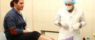
The night before hospitalization for surgical treatment, it is necessary to shave the sacrococcygeal and, if necessary, the gluteal region. It is possible to perform laser (Alexandrite or diode laser) or photoepilation a few days before hospitalization. However, the last two methods are ineffective for removing light hair. Another method of hair removal is electrolysis; it is more painful, but is suitable for all hair types. The effect after the procedures may not be achieved immediately, so it is better to perform them in advance - 14 days before the operation. Shaving, as an alternative to hair removal, will take you less time and money, but at the same time it may damage the skin, which can become a source of infection. No other special preparation is required for the operation; it will be enough to refuse food and liquids 8 hours before the operation.
Pilonidal cyst of the coccyx: diagnosis
Diagnosis of a pilonidal cyst is usually not difficult. When making a diagnosis, experienced proctologists at the Yusupov Hospital take into account the results of a visual examination of the area of the intergluteal fold, digital rectal examination and other instrumental studies, which are carried out using the latest diagnostic equipment of the clinic:
- sigmoidoscopy, which allows you to determine the location of the cyst with maximum accuracy;
- probing the cyst - to determine the canal of the coccygeal passage;
- ultrasound examination - to clarify the location and extent of the process;
- radiography of the sacrum with contrast;
- computer and magnetic resonance imaging.
CLINIC
As a result, patients report swelling of the perianal area, which, when inflamed, is accompanied by pain and palpation. Sometimes bleeding from the rectum is eliminated without pain. As a matter of fact, the patient may suffer from the consolidation of the inherited obstruction of the rectum or the mother may develop recurrent infections of the cervix of the inherited cervical obstruction. As Krizovo-Kuprikova teratoma directly affects the nerve pathways cauda equina
Or it metastasizes in the spinal cord, the patient may have maternal neurological scars, but it is even rare. With physical stimulation, similar perianal cysts will appear.
Brushes for anal passages/ducts.
As a matter of course, stench manifests itself as perianal pain, swelling and induration. These are spherical, smooth epidermal nodules, 1–2 cm in size. Most often, the stink is released in front of the anus.
Krizovo-Kuprikov's teratoma.
These drugs may be asymptomatic, but they may occasionally be detected during rectal examination. As a matter of fact, these soft tissues are localized presacral, ranging in size from 5 to 25 cm.
Dermoid brushes.
Typically, these are spherical rims with a size of 6–60 mm. The stench can be from the mother's sinus, which is what is called hair protruding. The problem is recurrent infection.
Epidermoid brushes.
These brushes are located near the dermis, and the stinking little bits lift the epidermis above themselves, creating thick, elastic dome-like structures with a yellowish-white color, which shifts within structures such as the hall. talk smarter. The stench can be found in the central canal, filled with keratin, and its diameter varies from several millimeters to 5 cm. More often than not, the stench is multiple. Over time, your brushes will become larger, and they may become inflamed and white.
Differential diagnosis must be carried out with pilonidal cysts, perianal abscesses, hemorrhoids and malignant tumors, hematomas due to trauma.
During diagnosis, pelvic computed tomography and transrectal ultrasonography provide valuable information. Endoscopic examination is required to exclude rectal cancer. In times of doubt, cytological examination can be taken instead of the brush removed by puncture.
Pilonidal cyst of the coccyx: treatment
Pilonidal cyst of the coccyx can only be treated surgically. During the operation, surgeons at the Yusupov Hospital eliminate the main source of inflammation - the epithelial canal, as well as the primary openings and surrounding tissues.
Surgical intervention for removal of a pilonidal cyst of the coccyx should be performed as planned after the patient's acute symptoms have subsided.
How to choose the right clinic and surgeon for treatment in my case?
To summarize the review of surgical treatment methods, it should be said that the choice of the type of operation is undoubtedly the prerogative of the surgeon, but today this decision is made together with the patient. When discussing the plan of surgical intervention (the scope of surgical intervention) specifically in your case, the surgeon should offer you various modern instruments (devices) necessary for the operation, while telling you the advantages and disadvantages of their use. If, in a conversation with you, the surgeon does not try to discuss different approaches to treating your specific ECC, but offers a non-alternative method, then this often indicates that there is no place for other methods in his arsenal.
In such a situation, you have the right to consult another doctor for a “second” opinion. It is necessary to be especially careful when choosing both a surgeon and an institution for surgical treatment in cases where there is a complex or recurrent ECC, when treatment often involves extensive excision of tissue in the sacral area.
Pilonidal cyst of the coccyx: postoperative period
Patients who have undergone radical surgery to remove a pilonidal cyst of the coccyx must follow a number of the following recommendations in the postoperative period:
- For three weeks after surgery, you should not take a sitting position; prolonged lying on your back and lifting heavy objects are also prohibited;
- after the stitches are removed, you need to take a shower, wash the intergluteal fold, wear clean underwear made from natural materials;
- avoid overheating and severe cooling;
- Six months after the operation, the patient should undergo hair removal in the coccyx area;
- Two to three months after surgery, it is recommended to stop wearing tight, tight clothing.
It is necessary to understand that early contact with a proctologist increases the likelihood of maintaining health and reducing the risk of developing unwanted complications.
The proctology department of the Yusupov Hospital provides high-quality services for the diagnosis and treatment of pilonidal cysts of the coccyx using the latest medical equipment from the world's leading manufacturers. High efficiency of treatment of pathology is ensured by the use of innovative technologies, as well as the extensive practical experience of proctologists at the Yusupov Hospital. Qualified rehabilitation specialists at the clinic develop an individual set of rehabilitation measures during the rehabilitation period after surgery for each patient. The Yusupov Hospital has created all the conditions necessary for a comfortable stay for patients within the hospital walls, and provides round-the-clock, high-quality care from medical personnel.
Who is at risk for developing ECC?
The disease occurs 4 times more often in men than women. ECC belongs to a group of rare diseases and is detected in only 26 out of 100,000 people. Mostly, young people of working age from 15 to 30 years old are affected. According to statistics, ECC most often occurs in Arabs and Caucasian peoples, less often in African Americans.
Risk factors for the development of ECC are:
- excess hair growth
- overweight
- insufficient attention to hygiene of the coccyx area
- passive lifestyle
- wearing tight and tight clothing (pants, skirts)
LIKUVANNYA
Injections of phenol can be widely used in the treatment of pilonidal sinuses in Europe, both in cases of acute abscesses and in chronic illness. Introduce 80% of the phenol into the sinus, add 1 quill there and then remove it. After this, the sinus is scraped with a curette. This procedure can be repeated three times, and repeated courses of bathing, if necessary, can be completed at an interval of 4–6 days. The skin near the sinus is treated with phenol, which is the epidermis, with paraffin gel.
Phenol sterilizes the sinus and removes hair, which injections can be combined with local sinus excision. As a matter of course, the wound will heal in 4–8 days. The relapse rate becomes 9–27% and is similar to that observed after simple hanging and packing of a closed wound. Due to the intense inflammatory reaction of patients, they are deprived of one dose from the hospital, and it is then recommended to take clean baths and shin pads.
Based on the choice of method of surgical treatment, pilonidal disease is divided into three groups: acute pilonidal abscess, chronic pilonidal disease, complex or recurrent pilonidal disease.
For pilonidal abscess
it is opened, curettage and drainage are performed. If it is possible to drain it through the incision on the side in the middle line, the fragments of the wound deep in the fold between the sits will burn badly. An important element to note is that the lower leg hair grows near the wound and scar for up to 3 months.
It is necessary to put pressure on the patient so that the opening of the abscess does not guarantee the absence of relapse. If curettage is not performed, in 60% of patients the wound will continue to heal for 2 months, but in 40% of them a relapse occurs. The leaders believe that up to 85% of the sick will require radical treatment in the near future. Extension of the pilonidal sinus canal reduces the relapse rate to 15%. It is often impossible to reveal fragments of this before the first delivery, so it is recommended to stop giving it a little again a week after opening the abscess, if the swelling subsides.
Concept of chronic pilonidal sinus
There are patients in whom there is a pilonidal drainage abscess, as well as patients in whom there is chronic sinus disease without the development of an acute abscess. There are many approaches to surgical treatment of such patients. The first one lies at the superior sinus and lateral sinuses, straightening the empty space to the presacral fascia with the removal of hair, granulation tissue and detritus of the gastrointestinal tract and behind the help of a curette. The skin hangs sparingly. The wound is treated with a second tension of 5–8 tension. Bascom, having opened the opening of the sinus and its epithelial canal from a small incision with the primary suture, and opened the empty one and sanitized from the lateral incision - such a wound will heal faster and require less dressings. The method of asymmetrical cuts is to change the interstellar fold and eliminate the frictional forces that act on the edges of the wound at the interstellar fold. The wound can also be closed by moving the skin cap.
Few techniques for applying the primary suture are effective in the presence of infection and permanently suspended emptying, with a high incidence of infectious complications, frequent inability to apply tension, and a high rate of relapses (up to 38%). Although closing an open wound with a second tension is more likely, there are fewer relapses of illness (8-21%). The lower frequency of relapses is associated with the creation of a wide, flat scar, visible from the hair follicles. This protects against skin friction, hair penetration into the skin and follicular infection.
A compromise between these approaches is marsupialization, in which the edges of the wounds after the hanging sinus are sutured to the presacral fascia, and the wound is packed.
The problem is the treatment of recurrent pilonidal disease
or wounds of a folding shape after hanging sinus. Relapses often result from regrowth of hair in the scar at the interstitial site, accumulation of sweat and infection in it. This problem is solved by the methods of plastic surgery: the wound, granulation and scar tissue are left hanging, after which the wound is sutured with the first suture, liquidating the interstitial fold with a stitch of stiffening method. ів plastics of the skin (movement of V or Y-like clamps of the skin or skin-meat claptiv). According to the data of various authors, the frequency of relapses can be reduced to 1.3–20%.
