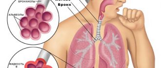
Community-acquired pneumonia is an infectious disease characterized by an inflammatory process in the lungs. The disease most often develops during influenza epidemics and acute respiratory viral infections. Patients with mild pneumonia can be treated on an outpatient basis. In cases of moderate to severe pneumonia, they are hospitalized in a therapy clinic.
The Yusupov Hospital employs professors and doctors of the highest category, who are leading experts in the field of pulmonology. To diagnose the disease, innovative examination methods are used to identify the pathogen, the localization and extent of the pathological process, and the severity of the disease. Doctors take an individual approach to the treatment of each patient, prescribing the most effective modern drugs with minimal side effects.
Causes of community-acquired pneumonia
Community-acquired pneumonia is conventionally divided into 3 groups:
- pneumonia that does not require hospitalization;
- pneumonia requiring hospitalization of patients in a hospital;
- pneumonia requiring hospitalization of patients in intensive care units.
The therapy clinic is staffed by experienced doctors and nurses. They treat each patient carefully. The rooms are equipped with air conditioning to ensure comfortable temperature conditions. Patients receive complete nutrition, rich in proteins and carbohydrates, and are provided with individual hyena products.
Patients with severe pneumonia are hospitalized in the intensive care unit. Resuscitation doctors monitor the functioning of the respiratory and cardiovascular systems around the clock and determine the level of oxygen saturation in the blood. If there are indications, artificial ventilation of the lungs is performed using stationary and portable expert-class devices.
The following types of community-acquired pneumonia are distinguished: pneumonia in patients without impaired immunity and pneumonia in patients with impaired immunity against the background of the advanced stage of AIDS or other diseases associated with impaired immune system.
Aspiration pneumonia develops in patients with swallowing disorders. Vomit can enter the respiratory tract during a disturbance of consciousness, stroke, acute traumatic brain injury, or during an epileptic attack.
Community-acquired pneumonia develops when microorganisms enter the respiratory tract. There are the following routes of infection for pneumonia:
- microaspiration of oropharyngeal contents;
- inhalation of aerosol containing microorganisms;
- hematogenous spread of microorganisms from foci of infection located outside the lungs;
- spread of infection from neighboring organs.
Pneumonia develops in the event of a weakening of specific or nonspecific protection, a large number of bacteria that have penetrated into the alveolar part of the lungs, or the penetration of microorganisms that are highly aggressive.
The most common causative agent of community-acquired pneumonia with a mild course is pneumococcus. Currently, great importance is attached to the occurrence of pneumonia in the presence and treatment of mycoplasma and chlamydia, Haemophilus influenzae and gram-negative enterobacteria, viruses and legionella. In patients with aspiration pneumonia, the more typical pathogens are gram-negative enterobacteria and anaerobic microflora.
Classification
Today, pneumonia is classified according to the conditions of the onset of the disease:
- community-acquired pneumonia (which a person became infected with not in the hospital, but at home, on the street, etc.)
- hospital/nosocomial
- pneumonia in people with immunodeficiency conditions
- aspiration
Based on severity, community-acquired pneumonia is divided into 3 groups:
- no need for hospitalization (mortality rate 1% to 5%)
— in which hospitalization of patients in a hospital is necessary (mortality rate is 12%)
— in which hospitalization of patients in the ICU is necessary (mortality rate is 40%)
Severe community-acquired pneumonia is pneumonia that has a high mortality rate and requires the patient to be admitted to the ICU. If a person has severe sepsis or septic shock, respiratory failure and the prevalence of pulmonary infiltrates according to an x-ray examination, then we are talking about severe community-acquired pneumonia.
Symptoms and diagnosis of community-acquired pneumonia
Pneumonia (community-acquired pneumonia) is manifested by clinical symptoms such as weakness, fatigue, nausea, lack of appetite, and impaired consciousness. Patients are bothered by a cough with sputum production and chest pain.
When examining a patient, the doctor may detect cyanosis, the lag of one half of the chest during breathing. During percussion, a shortening of the sound above the lesion is determined. During auscultation, weakened or bronchial breathing, crepitus, dry or moist rales are heard.
The diagnosis of pneumonia without radiological signs of pneumonia is invalid. Doctors at the Yusupov Hospital do x-rays of the lungs in two projections or large-frame fluorography. If the clinical picture does not correspond to radiological data, computed tomography is performed.
The purpose of microbiological research for community-acquired pneumonia is to isolate the pathogen from the source of infection. In a general blood test, the number of leukocytes and the erythrocyte sedimentation rate increase.
The diagnosis of pneumonia is considered established if the patient, against the background of an infiltrate in the lung tissue detected on an x-ray, has at least two clinical signs:
- acute onset of the disease with high body temperature;
- cough with sputum;
- physical signs of compaction of lung tissue;
- leukocytosis.
Acute pneumonia
The dominant role in the etiology of acute pneumonia belongs to infection, primarily bacterial. Typically, the causative agents of the disease are pneumococci (30-40%), mycoplasma (6-20%), Staphylococcus aureus (0.4-5%), Friedlander's bacillus, less often - hemolytic and non-hemolytic streptococcus, Pseudomonas aeruginosa and Haemophilus influenzae, fungi and their associations ; Among the viruses are influenza virus, RS virus, adenoviruses. Purely viral acute pneumonia is rare; usually ARVI facilitates the colonization of lung tissue by endogenous or, less commonly, exogenous bacterial microflora. In ornithosis, chicken pox, whooping cough, measles, brucellosis, anthrax, salmonellosis, the development of acute pneumonia is determined by the specific causative agent of this infection. Microorganisms enter the lower parts of the respiratory tract through the bronchogenic route, as well as hematogenous (in infectious diseases, sepsis) and lymphogenous (in case of chest injury) routes.
Acute pneumonia can occur after exposure to the respiratory tract of light chemical and physical agents (concentrated acids and alkalis, temperature, ionizing radiation), usually in combination with secondary bacterial infection with autogenous microflora from the pharynx and upper respiratory tract. Due to the long-term use of antibiotics in the development of acute pneumonia, the role of opportunistic microflora has become more significant. There are cases of allergic (eosinophilic) acute pneumonia caused by helminthiases and medications. Acute pneumonia can occur uncomplicated and with complications; mild, moderate or severe; with the absence or development of functional disorders.
Various factors that reduce the resistance of the macroorganism predispose to the occurrence of acute pneumonia: prolonged intoxication (including alcohol and nicotine), hypothermia and high humidity, concomitant chronic infections, respiratory allergies, nervous shock, infancy and old age, prolonged bed rest. The penetration of infection into the lungs is facilitated by impaired patency and drainage function of the bronchi, inhibition of the cough reflex, insufficiency of mucociliary clearance, defects in pulmonary surfactant, decreased local immunity, including phagocytic activity, levels of lysozyme and interferon.
In acute pneumonia, inflammation affects the alveoli, interalveolar septa and vascular bed of the lungs. Moreover, in different parts of the affected lung, different phases can be simultaneously observed - flushing, red and gray “hepatization”, resolution. Morphological changes in acute pneumonia vary depending on the type of pathogen. Some microorganisms (staphylococcus, Pseudomonas aeruginosa, streptococcus) secrete exotoxins that cause deep damage to the lung tissue with the appearance of multiple small, sometimes merging foci of abscess pneumonia. In acute Friedlander pneumonia, extensive infarct-like necrosis occurs in the lungs. Interstitial inflammation dominates in pneumonia of pneumocystis and cytomegalovirus origin.
Treatment and prevention of community-acquired pneumonia
Most patients with signs of community-acquired pneumonia can be treated on an outpatient basis. The antibiotic is chosen empirically, before receiving the results of a microbiological study, since any delay in antibiotic therapy for pneumonia is accompanied by an increased risk of complications.
The choice of initial therapy depends on the severity of the disease and clinical signs of pneumonia. To treat mild pneumonia on an outpatient basis, doctors prescribe oral amoxicillin and amoxicillin clavulanate. If pneumonia caused by atypical pathogens is suspected, oral macrolides or respiratory fluoroquinolones (levofloxacin, moxifloxacin) are used.
At the therapy clinic, pulmonologists prescribe comprehensive treatment for community-acquired pneumonia. Indications for parenteral antibiotic therapy are:
- disturbance of consciousness;
- severe pneumonia;
- violation of the swallowing reflex;
- functional or anatomical causes of impaired absorption.
For mild pneumonia, doctors use amoxicillin clavulanate, ampicillin, and parenteral cephalosporins of the second and third generations. Alternative drugs include intravenous macrolides or respiratory fluoroquinolones. If aspiration pneumonia is suspected, amoxicillin clavulanate or a combination of b-lactams with clindamycin or metronidazole is prescribed.
For severe pneumonia, a combination of third generation cephalosporins and macrolides is used. An alternative regimen is a combination of fluoroquinolones with third-generation cephalosporins. After receiving an adequate response to parenteral administration of antibacterial drugs, they switch to oral antibiotics.
Prevention of pneumonia includes a set of specific and nonspecific measures. To prevent pneumonia, it is necessary to observe a work and rest schedule, ventilate work and living areas several times a day for 30 minutes, regularly do wet cleaning, and eat well. It is recommended to play sports, do breathing exercises, quit smoking and alcohol abuse, and promptly sanitize foci of chronic infection. Vaccination against pneumococcal infection is carried out for older people with concomitant pathologies that increase the risk of developing the disease. For this, the Pneumo 23 vaccine (made in France) is used.
Forecast
The mortality rate of patients with severe community-acquired pneumonia hospitalized in the ICU is high and ranges from 22 to 54%. An unfavorable prognosis may be due to the following factors:
- performing mechanical ventilation
- patient age 70 years or older
- bacteremia
- bilateral localization of pneumonia
- need for inotropic support
- sepsis
- P. aeruginosa infection
- ineffectiveness of initial (initial) antibiotic therapy
The fatality rate is high when community-acquired pneumonia is caused by Legionella spp., S. pneumoniae, P. aeruginosa, or Klebsiella pneumoniae.
Laboratory and instrumental diagnostics
Leukopenia below 3x10⁹/l or leukocytosis above 25x109/l is considered an unfavorable prognostic sign of septic complications.
The following changes are possible in the patient's blood test:
- Leukocytosis or leukopenia
- Neutrophil shift in leukocyte formula
- Increasing ESR
- Increased acute phase reactants (CRP, fibrinogen, procalcitonin test). Other changes in the biochemical blood test may indicate decompensation in other organs and systems
Determination of arterial blood gases/pulse oximetry is performed in patients with signs of respiratory failure, massive effusion, and the development of CAP due to chronic obstructive pulmonary disease (COPD). Decrease in PaO2 below 60 mm Hg. Art. is an unfavorable prognostic sign and indicates the need to admit the patient to the intensive care unit (ICU).
A bacterioscopy of the patient's sputum is carried out to identify the pathogen, and the sputum is cultured on nutrient media to determine sensitivity to antibiotics. For severe patients, it is recommended to collect venous blood for blood cultures (2 blood samples from 2 different veins, sample volume - at least 10 ml), PCR study, serological diagnostics to identify respiratory viruses, atypical pathogens of bacterial pneumonia.
X-ray examination of the lungs usually reveals focal infiltrative changes and pleural effusion. Chest CT may be a useful alternative to radiography in a number of situations:
- In the presence of obvious clinical symptoms of pneumonia and no changes on the radiograph
- In cases where, during examination of a patient with suspected pneumonia, atypical changes are revealed (obstructive atelectasis, pulmonary infarction due to pulmonary embolism, lung abscess, etc.)
- Recurrent pneumonia, in which infiltrative changes occur in the same lobe (segment), or prolonged pneumonia, in which the duration of existence of infiltrative changes in the lung tissue exceeds 4 weeks
Fiberoptic bronchoscopy with a quantitative assessment of microbial contamination of the obtained material or other invasive diagnostics (transtracheal aspiration, transthoracic biopsy) is performed if pulmonary tuberculosis is suspected in the absence of a productive cough, as well as with bronchial obstruction by bronchogenic carcinoma or a foreign body
If the patient has pleural effusion and there are conditions for safe pleural puncture, it is necessary to conduct a study of the pleural fluid: counting leukocytes with a leukocyte formula; determination of pH, lactate dehydrogenase activity, protein content; Gram staining of smears; culture to identify aerobes, anaerobes and mycobacteria.
Treatment
It is fundamentally important to determine the patient’s management tactics - whether he should be hospitalized or whether treatment can be carried out on an outpatient basis. For these purposes, you can use the CURB-65/CRB-65 scale, which involves assessing a number of indicators (presence - 1 point, absence - 0):
C —impaired consciousness
U - blood urea nitrogen more than 7 mmol/l (not taken into account in CRB-65)
R — respiratory rate ≥ 30 per minute.
B - systolic blood pressure < 90 mm Hg or diastolic blood pressure < 60 mm Hg. Art.
65 - age 65 years and older
Explanation:
0 points - the patient belongs to group I (mortality 1.2%) and is treated on an outpatient basis.
1–2 points correspond to group II (mortality 8.15%), patients in this group are subject to hospitalization with observation and dynamic assessment of the condition.
3 points and above - patients are subject to emergency hospitalization; mortality in this group can reach 31%.


