Author:
Amelicheva Alena Aleksandrovna medical editor
Quick Transition Treatment of Amyopathic Dermatomyositis
Amyopathic dermatomyositis is a form of dermatomyositis, a rare disease belonging to the group of inflammatory myopathies.
In amyopathic dermatomyositis, inflammatory and degenerative skin changes characteristic of dermatomyositis are observed. But, unlike dermatomyositis, there are no signs of damage to skeletal and smooth muscles (progressive symmetrical muscle weakness of the proximal limbs).
Amyopathic dermatomyositis affects women more often than men (3:1). The onset (first clinical manifestation) of the disease is usually observed at the age of 20-40 years.
Causes
The exact causes of amyopathic dermatomyositis have not yet been established. It is assumed that the triggers of the disease may be immune, genetic and environmental factors. Amyopathic dermatomyositis is classified as an autoimmune disorder because patients with this diagnosis experience abnormal production of autoantibodies by the immune system, leading to inflammatory and degenerative changes in the skin.
It is also possible that the root cause of the development of pathology may be infections (Coxsackie virus, parvoviruses, HIV, echoviruses, Toxoplasma, Borrelia), malignant neoplasms, and certain medications (statins, penicillamine, phenylbutazone). However, these data are still probabilistic statements and are still being studied.
General information
Dermatomyositis , or in other words, Wagner's disease , is a severe diffuse progressive systemic disease of connective tissue that affects skeletal and smooth muscles, the microcirculatory system of blood vessels, internal organs and skin.
Leads to the replacement of functional structures with fibrous ones. The clinical picture is expressed in the form of motor dysfunction, vasculitis , edema, erythema , hyperkeratosis and may be:
- acute , starting with fever and a memorable development of symptoms, without treatment leads to death after 2-3 months, caused by complete immobilization and severe cardiopulmonary failure;
- subacute - with a gradual increase over several years of muscle weakness, arthralgia , erythema, changes in internal organs;
- chronic - with a benign outcome, having a cyclical course and long-term remissions, characterized by the presence of local atrophic and sclerotic changes in the muscles and skin, and compensated changes occur in the internal organs.
As a result, dermatopolymyositis leads to complications - fibrosis, calcification , atrophy and purulent infections.
Wagner's disease is rare - 2-6 adults and 1-2 children per 100 thousand people. The pathology is more common in countries in southern Europe. The incidence increases in the spring-summer season, which is associated with insolation - more intense exposure to sunlight.
Symptoms
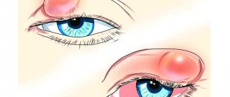
- Heliotropic rash (erymatous spots with a purple tint in the periorbital area, around the eyes) in combination with edema;
- Gottron's papules (nodules and plaques of red and purple color on the interphalangeal and phalangeal joints);
- Gottron's symptoms (erymatous rash with a purple tint, affecting the extensor surface of the elbow and knee joints);
- itching, dryness, peeling on the areas of the skin affected by the rash;
- peeling, cracks on the skin of the fingers and palms on the inside;
- erythema (redness) of the scalp;
- poikiloderma (dystrophic changes in the skin with telangiectasia, areas of atrophy, hypo- and hyperpigmentation);
- photosensitivity;
- hair thinning;
- redness of the periungual fold, growth of skin around the nail bed.
Classification
In approximately 25-30% of cases, there are no skin symptoms and then they speak of a special case of this systemic disease - polymyositis .
Depending on the origin and course, the following are distinguished:
- primary idiopathic dermatopolymyositis;
- the secondary paraneoplastic or tumor type occurs in individuals over 55 years of age against the background of cancer of the breast , lungs , ovaries , prostate , uterus , stomach , colon , hemoblastosis and other malignant neoplasms;
- juvenile - children's;
- polymyositis , combined with other diffuse connective tissue diseases.
Juvenile dermatomyositis: features of the course
It is distinguished by focal calcification of soft tissues such as muscles, epidermis and subcutaneous fat. Deposits of calcium salts - hydroxyapatites - can be deposited on the surface and deep, forming limited single or diffusely located nodules and pseudotumors. Superficial plaques with calcifications cause inflammatory reactions of surrounding tissues; they rot and are rejected in the form of a crumbly mass.
Subcutaneous calcifications
As in adults, classic rashes and Gottron's sign are observed. Gradually increasing muscle weakness begins with a bizarre gait of the child, which further constrains movement even more, preventing the child from playing, running and caring for himself.
Juvenile dermatomyositis
Diagnostics
The doctor interviews the patient, conducts a physical examination, collects a complete history of the disease, and gives directions for the necessary studies.
The diagnosis of amyopathic dermatomyositis is established for patients in whom skin lesions characteristic of dermatomyositis are confirmed by biopsy results. However, laboratory and instrumental diagnostic methods did not reveal clinical signs of muscle tissue damage.
Additionally, such patients may be tested for the presence of malignant neoplasms (dermatomyositis is associated with cancer) or interstitial lung disease.
Diet for dermatomyositis
Vitamin-protein diet
- Efficiency: 5 kg in 7-10 days
- Terms: up to 14 days
- Cost of products: 1700-1800 rubles. in Week
Since the pathology can affect the muscles of the pharynx and cause dysphagia, patients are recommended to eat pureed and liquid food. The diet should contain:
- fresh juices;
- vegetable cream soups and purees;
- light dietary protein in sufficient quantities.
Moreover, it is important to maintain normal proportions of BJU and use natural products, as well as healthy methods of processing them.
Treatment of amyopathic dermatomyositis
Treatment of amyopathic dermatomyositis is aimed at controlling skin manifestations. The first line of therapy includes protection from sun exposure (use of sunscreens), as well as the use of topical steroids and antihistamines aimed at reducing erythema and itching. If treatment fails, oral corticosteroids and antihistamines are prescribed.
If there is no positive effect, the patient may be prescribed injections of corticosteroids, antimalarial (hydroxychloroquine) or cytostatic (methotrexate) drugs, immunosuppressants (mycophenolate), and intravenous immunoglobulin.
Patients diagnosed with amyopathic dermatomyositis require constant medical supervision and health monitoring to promptly identify signs of muscle tissue inflammation.
Features and advantages of treatment of amyopathic dermatomyositis at the Rassvet clinic
The Rassvet clinic employs a multidisciplinary team of highly qualified specialists in all areas. All our doctors are well trained and have extensive practical experience necessary to diagnose and treat not only typical diseases, but also rare diseases and syndromes.
Patients with suspected amyopathic dermatomyositis undergo all necessary studies, are prescribed therapy aimed at reducing the symptoms of the disease and improving the patient’s quality of life, and provide maximum support at all stages of treatment.
Pathogenesis
The pathogenetic process begins with a nonspecific effect - hypothermia, excessive solar insolation , vaccination , acute intoxication , trauma and other exogenous factors. It is associated with the presence of infectious agents introduced into the genome of muscle cells, changes in neuroendocrine reactivity or genetic predisposition - the presence of HLA histocompatibility antigens associated with B8 or DR3 .
With dermatomyositis, an immune-inflammatory reaction develops, aimed at the destruction of altered intranuclear structures of muscle and skin cells. Due to cross-reactions, cell populations related to antigens are involved in immune damage. Microphage mechanisms of elimination of immune complexes from the body lead to activation of fibrogenesis and systemic inflammation of small vessels. The hyperreactivity of the immune system is aimed at destroying the intranuclear structure of the virion; it is manifested by the presence in the bloodstream of antibodies Mi2, Jo1, SRP, as well as autoantibodies to nuclear proteins and other soluble antigens.
Photos of dermatomyositis and polymyositis
Polymyositis and dermatomyositis are rheumatic diseases, the features of which are the appearance of inflammation and transformation of muscles (polymyositis) or muscles and skin. A more characteristic dermatological sign is heliotrope rash. If you have any similar symptoms listed above, you must definitely contact the clinic to rule out the occurrence of the disease. Below are photos of dermatomyositis on different parts of the body.
Treatment with folk remedies
Dermatomyositis is difficult to treat, but with patience, results can be achieved using folk remedies.
You need to understand that plant treatment is used during the period of decreased signs and pronounced symptoms. Treatment is carried out in the spring and autumn to prevent exacerbation. The course of treatment lasts for a month.
Let's consider traditional methods of treatment:
- Treatment consists of applying compresses and lotions. To prepare a compress, you need ingredients such as willow leaves and buds (1 tbsp each). All components are filled with water and brewed. After cooling, you can apply it to painful areas of the body.
- You can also use the following recipe and make lotions: take marshmallow (1 tbsp) and pour a glass of boiling water over it, brew it.
- To prepare ointments you will need willow and butter. After preparation, the medicine can be applied to the affected areas.
- The following composition of ingredients helps perfectly with the disease of dermatomyositis: oats (500 g), milk (liter or one and a half). Place the purchased mixture on low heat and cook for two minutes. After the tincture has cooled, it must be strained. Treatment lasts for a month, you can drink up to a glass of decoction a day.
Prognosis and prevention
Today, thanks to the use of effective medications, the development of dermatomyositis disease is inhibited, and with the supervision of a qualified doctor, improvement occurs quickly.
So, when the doctor has prescribed the exact dosage of the medicine, you do not need to reduce the amount of the drug yourself. It is precisely because of the reduction in dosage that the patient’s situation worsens.
Dermatomyositis in the protracted stage of the disease, despite therapy, has a high probability of complications.
The earlier the diagnosis is made and treatment prescribed, the higher the likelihood of a complete recovery for the patient. The child may also end up with absolute recovery or stable remission.
Measures that would prevent the formation of the disease have not been created to date. However, in clinics such preventive measures include the following actions:
- maintenance drug therapy;
- periodic examinations with doctors, especially a dermatologist and rheumatologist;
- conducting tests to exclude tumors;
- treatment of inflammatory diseases;
- getting rid of the sources of the infectious process in the body.
Illness in children
Juvenile (childhood) dermatomyositis is characterized by symptoms of muscle inflammation and weakness, which subsequently leads to limitation of physical activity. A characteristic feature of the disease in children, which distinguishes it from adult dermatomyositis, is the formation of the disease without the occurrence of tumors.
The causes of its appearance in children are often considered in terms of the influence of infections. There is an opinion that the disease of dermatomyositis at a young age is due to heredity. Irradiation of surfaces by sunlight is of great importance in the formation of the disease. The main signs of childhood (juvenile) dermatomyositis include muscle inflammation, muscle weakness, dermatological rashes, skin diseases, diseases of the lungs and intestinal tract.
As a result of the extremely rapid spread of the disease in the children's body, children die more often than adults. Deaths have been recorded already during the first years of the disease. Naturally, if you approach the treatment process competently and throughout the disease follow the doctor’s recommendations, take the necessary medications and improve muscle function in every possible way, then the disease can be overcome. On average, treatment lasts for three years, but in some cases – up to 15 years.
