Cholangitis is an inflammation of the bile ducts, occurring in acute or chronic form. In most cases, it occurs due to the penetration of a bacterial or parasitic infection from the intestines, gall bladder or through the circulatory system. In this case, the patient’s skin and sclera of the eyes turn yellow, pain appears in the hypochondrium on the right side, and the temperature rises greatly.
The risk of this disease increases with age; most often the disease occurs in women between 50 and 60 years of age. The available statistics are due to the fact that by advanced age, people accumulate a number of past or untreated diseases (for example, gastritis, cholelithiasis, duodenitis, hepatitis, etc.), which can cause complications in the form of cholangitis.
In the international classification of diseases created by WHO (World Health Organization) to standardize medical diagnoses, various forms of cholangitis appear under several headings. The main disease code according to ICD-10 is K83.0.
Classification
The course of the disease is:
- Acute (occurs unexpectedly).
- Chronic (appears after an acute condition, either as an independent disease or as a result of complications of diseases of the digestive system).
Acute cholangitis is divided into:
- Purulent. With this pathogenesis, the liver and gallbladder are also susceptible to inflammation, since purulent cholangitis is characterized by the filling of the bile ducts with pus.
- Catarrhal. It manifests itself as swelling of the bile ducts, often ending in the formation of scars and narrowing of the ducts.
- Necrotic. It occurs as a result of pancreatic enzymes entering the bile ducts, which is accompanied by tissue death.
- Diphtheritic. The formation of ulcers and necrosis in the bile ducts leads to rotting of the walls and connective tissues of the ducts. In this case, the disease can affect neighboring organs.
Chronic cholangitis is divided into:
- Sclerosing, or, as it is also called, sclerosing cholangitis. The connective tissue in the bile ducts becomes much larger than the normal amount, resulting in narrowing of the lumen and deformation. Such growth leads to stagnation of bile and the manifestation of other related diseases.
Today, primary sclerosing cholangitis is a liver disease of unknown etiology, i.e. the causes and conditions for the appearance of new tissues are not obvious. In this regard, leading hepatology organizations around the world have established a generally accepted format and are constantly updating clinical guidelines that outline the latest methods for diagnosing and treating the disease.
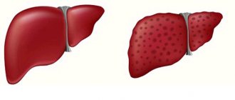
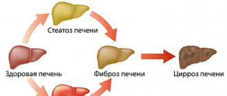
PSC (primary sclerosing cholangitis) is classified as an autoimmune liver disease, including primary biliary cholangitis (liver cirrhosis) and hepatitis. Often the patient experiences cross-syndromes of these diseases.
The secondary form of SC (sclerosing cholangitis) is caused by inflammatory processes of large and small pathways located outside and inside the liver. Most often, this condition occurs as a complication of choledocholithiasis or cholangiocarcinoma, due to injury or infection of adjacent organs.
- Recurrent. The symptoms of this condition are called “Charcot’s triad”. This means that the patient has a whole combination of symptoms of multiple sclerosis, fever, jaundice, as well as pain in the upper abdomen.
- Latent. The most difficult to diagnose option, in which symptoms appear barely noticeable or are absent at all. The only signs of the disease can be itchy skin, weakness and gradual enlargement of the liver. Listen to your body and if you experience similar reactions, immediately contact qualified specialists.
- Septic lingering. A severe form of the disease, manifested, among other things, by an enlargement of the spleen, liver, and inflammation of the kidneys.
- Abscessing. This inflammation is characterized by the formation of abscesses followed by necrosis.
1.What is cholangitis and its causes?
Cholangitis is a disease in which the bile ducts become inflamed. The intrahepatic bile ducts are a special system that distributes bile within the liver in both lobes. The right and left branches of the ducts, leaving the liver, form the hepatic duct. The hepatic duct, in turn, connects with the cystic duct, which drains bile from the gallbladder, to form the common bile duct - this is the scientific name for the common bile duct.
Causes of cholangitis
Sclerosing cholangitis is usually divided into primary, chronic and acute. Primary cholangitis is very rare in medical practice: more often cholangitis occurs as a result of cholelithiasis, pancreatitis, cholecystitis, tumors and abnormal development of the bile ducts, as well as after operations on the internal organs of the abdominal cavity.
Scarring of tissue, damage to the mucous membrane and stagnation of bile contribute to the development of the disease. In such cases, a bacterial infection or the appearance of parasites (for example, Giardia) can become a trigger for chronic or acute cholangitis.
Primary sclerosing cholangitis is not associated with infections, but develops as a result of autoimmune inflammation. As a result of inflammation, a narrowing of the intrahepatic and extrahepatic external ducts occurs. The causes of autoimmune inflammation are still unknown, but genetics as well as stress have been suggested to play a role.
A must read! Help with treatment and hospitalization!
Stages and symptoms of cholangitis in adults
| Stage | Symptoms |
| I | - severe chills due to a sharp increase in body temperature, occurring once every 1-3 days; - weakness; - low blood pressure; - vomit; - pain in the upper abdomen on the right. |
| II | The above symptoms plus: - increased size and pain of the liver; - yellow skin tone (jaundice); - increase in the size of the spleen. |
| III | The worsening of the condition is characterized by: - renal failure; - increased urea and creatine in the blood; - disturbances in the functioning of the heart (arrhythmia, tachycardia, muffled groans); - development of pancreatitis. |
| IV | At the last stage of the disease there is: - more severe hepatic-renal failure; - coma. |
In the table presented, the signs of inflammation of the bile ducts are summarized and can simultaneously be suitable for different variants of the course of the disease, therefore, if you find similar manifestations in yourself, we recommend that you consult a doctor for a full diagnosis of the disease.
Causes of the disease
When identifying the etiology of cholangitis, the main ways of spreading the infection are identified: lymphogenous (with inflammation of the small intestine, gallbladder or pancreas), hematogenous (in other words, with blood) and migration from the duodenum.
List of bacterial infections - causative agents of inflammation:
- staphylococcus;
- Proteus;
- pale spirochete;
- typhoid bacillus;
- enterococcus;
- coli.
In the case of a parasitic infection, helminths (worms) settle in the bile ducts, the long stay of which contributes to the formation of harmful bacteria.
Cholangitis can also develop against the background of the following ailments:
- stagnation of bile;
- kinks of the bile ducts;
- toxoplasmosis;
- ulcerative colitis;
- choledocholithiasis;
- Crohn's disease;
- biliary tract cancer;
- rheumatoid arthritis;
- cysts in the common bile duct;
- vasculitis;
- thyroiditis, etc.
The reason for the development of the disease in question can also be surgical intervention in adjacent organs, as a result of which the bile canaliculi were affected and damaged.
Treatment
The main goals of therapy are to relieve symptoms, stop the progression of the disease, prevent or eliminate complications of cholangitis, and optimize the time for liver transplantation.
Medication
. Cupruretic drugs and agents that prevent the development of fibrosis can be used in treatment. Immunosuppressive therapy may also be prescribed. The doctor selects specific medications based on the patient’s condition and medical history.
Cholangioplasty
. This is an operation to remove scar narrowing of the bile ducts. The use of this method can help reduce the severity of jaundice and relieve bacterial cholangitis. However, the procedure does not slow the progression of the disease.
Surgery
. For cholangitis, in addition to reconstructive operations on the biliary tract, proctocolectomy (removal of the colon and rectum) and liver transplantation can be performed.
Symptom relief
. To improve the quality of life, the patient may be prescribed medications to relieve itching. If there is a deficiency of fat-soluble vitamins, replacement therapy may be indicated.
Diagnosis of cholangitis
If symptoms indicating problems in the bile ducts are identified, the gastroenterologist prescribes a series of tests for the patient.
Stage I. Laboratory research
Includes analysis of patient biomaterials:
- Blood sampling for ESR (erythrocyte sedimentation rate), leukocytes and neutrophils (from a finger).
- General urine analysis.
- Biochemical blood test (from a vein).
- Immunological analysis of blood serum. It is performed if primary sclerosing cholangitis is suspected.
Stage II. Instrumental examination
This type of study will allow you to classify the disease and, in accordance with the data obtained, begin treatment.
The analysis is carried out using devices:
- MRCP (magnetic resonance cholangiopancreatography) is the main method for checking the bile ducts. It is based on the introduction of a special substance into the blood, which “highlights” the ducts during MRI scanning.
- Ultrasound (ultrasound examination) of the abdominal cavity.
- ERCP (endoscopic retrograde cholangiopancreatography) is used for diagnosis as the main type of examination in the absence of an MRCP (magnetic resonance cholangiopancreatography) apparatus. Unlike the first method, in this embodiment, the contrast agent is directly introduced into the duct system using a special device - a fibrogastroduodenoscope.
- PTC (percutaneous transhepatic cholangiography). The name of the method speaks for itself. A needle with a dye is inserted into the patient through the skin and liver. Then an x-ray is taken.
Based on the examinations performed, the attending physician begins to formulate a pathological diagnosis of cholangitis over time, taking into account the relationship with underlying diseases. As a result, the patient receives a detailed report.
Endoscopic diagnosis and treatment of acute cholangitis
Sokolov A. A., Professor of the Department of General Surgery of the Russian National Research Medical University named after. N. I. Pirogova Laberko L. A., professor, acting. O. head Department of General Surgery L/F RNRMU named after. N. I. Pirogova Lopatkin D. S., doctor of the endoscopy department of City Clinical Hospital No. 13 Moscow
Acute cholangitis is the most severe complication of various diseases of the bile ducts. The combination of obstructive jaundice with purulent cholangitis is observed in 20–30% of cases, while postoperative mortality in patients with this pathology is 15–45%. The main cause of mortality is progressive liver failure, and the development of cholangiogenic purulent complications aggravates the course of the disease and increases mortality. Despite the fact that purulent cholangitis is a companion to bile duct obstruction, it has now acquired the status of an independent problem.
To recognize the pathology of the extrahepatic bile ducts and cholangitis, the clinic uses clinical laboratory and special instrumental diagnostic methods (comprehensive abdominal ultrasound, computed tomography, magnetic resonance imaging, etc.).
However, the most complete and accurate information about the nature and level of obstruction to the outflow of bile was obtained using endoscopic retrograde cholangiopancreatography (ERCP).
Currently, this method is the most informative in diagnosing bile duct pathology; in addition, it allows performing various endobiliary interventions, which makes this method indispensable in the clinic.
To assess the condition of the bile ducts, ERCP with intervention on the BDS was performed in 1279 patients with obstructive jaundice. In 762 (59.6%) of 1279 patients, ERCP revealed choledocholithiasis, which in 33.4% of cases was multiple.
The size of the stones ranged from 0.5 to 3.5 cm. In 112 (8.8%) cases, choledocholithiasis was combined with stenosis of the common bile duct, in 28 (2.2%) - with stenosis of the terminal common bile duct (TC). In 41 (3.2%) cases, isolated stenosis of the BJ or TOX was diagnosed. Adenomatosis of the orifice of the BDS was diagnosed in 45 (3.5%) patients, tumor lesions of the BDS and extrahepatic bile ducts were diagnosed in 178 (13.9%) patients. In 76 (5.9%) cases, no pathology of the bile ducts was detected.
Particular difficulties in diagnosing acute cholangitis arose in the subclinical form of this pathology, when the clinical picture and laboratory data were absent or not expressed. In such cases, during ERCP, bile was collected from the common bile duct to determine the degree of bacterial contamination (bacteriocholia). Assessing the state of ductal bile parameters was important in determining the optimal tactics for drainage and sanitation of the bile ducts. The state of bile determines the characteristics of bile duct diseases, the course and prognosis of endogenous intoxication and postoperative complications. In 85% of cases in patients without clinical signs of acute cholangitis, the degree of bacteriocholia was > 10 5 microorganisms in 1 ml of bile, which is an indicator of the implementation of an active microbial inflammatory process in the biliary tract.
A comprehensive examination of a patient with obstructive jaundice syndrome in the preoperative period allows us to make an accurate diagnosis in a short time, determine rational treatment tactics for patients and set reasoned indications for endoscopic and surgical treatment of patients.
Of primary importance in the treatment of acute cholangitis is decompression of the bile ducts followed by their sanitation.
After decompression, tissue hemoperfusion of the liver and its excretory function improve, basal blood flow through the portal vein increases, the functional reserve of the liver increases, the increased flow of endotoxin into the bloodstream stops, which creates optimal conditions for restoring impaired functions of the liver and other organs and systems.
Endoscopic papillosphincterotomy (EPST) is the method of choice for patients with calculous inflammatory diseases of the pancreaticobiliary zone. The introduction of EPST into clinical practice has fully resolved the issues of decompression of the biliary system for a benign cause of obstruction, especially in patients with high surgical and anesthetic risk.
EPST was performed in 1124 (87.9%) cases. In the vast majority of cases (83.6%), it was performed using a typical cannulation method, while in the rest, non-cannulation or combined techniques were used. In 179 (15.9%) patients, the intervention was performed in two or more stages, which was due to technical difficulties or bleeding from the papillotomy incision. In 38.6% of cases, ERCP with intervention on the ampulla was of an emergency nature, which was associated with an increase in the phenomena of endotoxemia due to obstructive jaundice and purulent cholangitis, as well as when the syndrome of acute occlusion of the ampulla of the ampulla was detected. A high level of hyperbilirubinemia was not a reason to refuse decompressive intervention. Proper preoperative preparation based on blood coagulation parameters made it possible to prevent the development of severe postpapillotomy bleeding.
Endoscopic tactics for choledocholithiasis have now become more active due to the development of various methods of lithoextraction and lithotripsy. Lithoextraction is indicated for all patients, as it eliminates the need for repeated examinations, and there is no danger of wedging of unremoved stones into the TOC or ampoule of the BDS. In our observations, stones were removed simultaneously in 86.3% of cases; in other cases, their extraction required repeated manipulations.
For large common bile duct stones, various types of lithotripsy are used, each of them has its own advantages and disadvantages. The most widely used technique in the clinic is intraductal mechanical lithotripsy (IMLT). It is not entirely correct to determine indications for performing VMLT depending only on the size of the stone, since it is known that in some cases it is possible to remove stones with a diameter of 15 mm or more without destroying it, and in others, the removal of stones of a smaller size presents significant difficulties. In this regard, the success of lithoextraction depends not only on the size of the stone, but also on the size of the papillotomy opening. The latter, in turn, depends on the diameter of the intramural portion of the common bile duct. Indications for VMLT were given in 78 patients with stones ranging in size from 1.5 to 3.5 cm. This manipulation was effective in 71 (91.0%) cases.
An undoubted success in the treatment of acute cholangitis was the introduction into clinical practice of nasobiliary drainage (NBD) and endoscopic stenting of the bile ducts. External biliary drainage serves both for decompression, fractional or drip lavage of the biliary tract with antiseptics, and for the prevention of strangulation of stones in the gastrointestinal tract. In addition, external drainage makes it possible to perform an X-ray contrast study to monitor the passage of small or fragments of destroyed stones after VMLT.
In 118 cases, we included nasobiliary drainage and endoprosthetics of the common bile duct into the complex of endoscopic treatment. In 64.8% of cases, these methods were used to relieve the symptoms of purulent cholangitis. In other cases, external and internal biliary drainage was used to prevent the herniation of stones left in the common bile duct and to normalize the passage of bile into the intestine during tissue edema in the EPST area.
Treatment of acute cholangitis in patients with obstructive jaundice by systemic administration of antibacterial drugs is ineffective due to a violation of the absorptive-excretory function of hepatocytes against the background of a decrease in microcirculation in the liver caused by prolonged biliary hypertension. A more effective method is the local treatment of acute cholangitis. For these purposes, in addition to antiseptic solutions, we used instillation of an ozonized solution into the bile ducts through the NBD. This ensures the suppression of infection, improves the rheology of bile and its passage into the duodenum, due to the high bactericidal activity, local oxidative and biostimulating effect of ozone. However, we should not forget that cholestasis causes disturbances in the digestive, absorption, detoxification, barrier and other functions of the intestine, including due to the entry of highly toxic bile into its lumen. Therefore, the volume of liquid for sanitation of the biliary tract through the NBD was limited by us to 40–60 ml of antiseptic solution. In cases where the ducts were washed during control duodenoscopy using a catheter, the amount of antiseptic solution was increased to 100 ml, due to the fact that toxic bile entering the intestine was actively removed using suction. In 52 patients with nasobiliary drainage, local treatment of purulent cholangitis was carried out with freshly prepared ozonated saline solution, immediately after intervention on the obstructive system and for 3–5 days after it (once a day). The effectiveness of treatment was monitored based on clinical and laboratory data and the dynamics of bacteriocholia. Comparing the results of local treatment of purulent cholangitis with antiseptic solutions and ozonized saline, it was found that bacterial contamination of bile decreased to a level of less than 10 5 in 1 ml of bile in patients after local ozone therapy, faster by an average of 20.5 days than with traditional methods of sanitation .
Thus, the use of modern endoscopic methods for treating patients with obstructive jaundice syndrome and acute cholangitis due to biliary obstruction of various origins has increased the therapeutic effectiveness of the complex to 97.7%, reduced the number of complications from 10.3% to 7.5%, and decreased mortality — from 4.2% to 2.9%.
Treatment of cholangitis in adults
Depending on the extent and causes of the disease, treatment can be conservative or surgical.
| Basics of conservative treatment | Surgical operations |
|
|
Note! Advanced stages of the inflammatory process under consideration cannot be treated conservatively, because the ducts of the system are severely deformed.
Prognosis and prevention
The prognosis of the disease depends on many factors, ranging from the patient’s age to previous diseases and characteristics of the body.
Clearly, inflammation of the biliary tract is a serious disease that requires complete diagnosis and treatment. It is worth remembering that an incompletely cured disease will develop into a chronic form, in which the outcome is often unsatisfactory, since recovery is extremely difficult to achieve.
It is strongly not recommended to self-medicate, even with mild catarrhal form. Experienced doctors will be able to prescribe effective treatment and achieve a positive result in a short time.
The best remedy against cholangitis is prevention, because preventing the disease is easier than eliminating its consequences.
The following actions are recommended for those who have not encountered this problem or do not want to provoke relapses:
- Healthy food;
- maintain personal hygiene;
- walk in the fresh air more often;
- engage in physical education;
- periodically undergo examination by a gastroenterologist;
- promptly treat diseases of the gastrointestinal tract, liver and kidneys.

