Kaposi's sarcoma, what is it? First symptoms and methods of treatment
Kaposi's sarcoma in HIV-infected patients is one of the “AIDS indicator” diseases. The detection of a tumor of this type in young people without obvious disturbances in the functioning of the immune system provides grounds for diagnosing HIV infection in the AIDS stage, even without the use of laboratory research methods.
Kaposi's sarcoma accounts for 85% of all tumors developing in AIDS patients. This is a malignant multifocal tumor of vascular origin that affects the skin, mucous membranes and internal organs. It has several varieties, one of which is epidemic sarcoma associated with AIDS.
The tumor affects people under the age of 35 - 40 years, most often occurs in passive homosexuals, manifests itself as spots, nodules or plaques of a bright red or red-brown color, quickly spreads throughout all the skin, mucous membranes and internal organs. Elements of the tumor merge over time to form tumor-like formations, which eventually ulcerate. Kaposi's sarcoma is difficult to treat and quickly leads to death in patients. The correct diagnosis is easily established and confirmed by examining a piece of tissue under a microscope.
Causes
The reasons that cause the appearance of such neoplasms are not known for certain. But scientists with a high degree of probability suggest that the disease can develop against the background of human herpes virus type 8, which itself has not yet been sufficiently studied.
Kaposi's sarcoma also often accompanies other malignant processes, including:
- lymphosarcoma;
- multiple myeloma;
- mycosis fungoides;
- Hodgkin's lymphoma (lymphogranulomatosis);
- leukemia.
For pathology to occur, a significant decrease in human immunity due to various reasons is necessary. In addition, some groups of people have a much higher risk of developing Kaposi's sarcoma than others. For example, the disease is more often observed in men than in women.
At risk are:
- HIV-infected persons;
- persons with organ transplants from donors (especially kidneys);
- elderly men belonging to the Mediterranean race;
- persons whose homeland is equatorial Africa.
Medical scientists are unanimous on one thing: most often, especially in the initial stages of development, this disease is more likely to be a reactive process (that is, occurring in response to an infectious lesion) than a true sarcoma.
What chemotherapy regimens are used?
At the first stage, the most effective cytostatics with a lower likelihood of severe complications are used - these are anthracycline antibiotics, doxorubicin or daunorubicin, enclosed in special microcapsules. For tumors sensitive to cytostatics, 6 courses with administrations every two to three weeks are sufficient.
Both drugs act aggressively on the myocardium and blood cells, but due to the liposomal form, these side reactions are less pronounced than with conventional forms of cytostatics.
If the first line is ineffective - the continued growth of sarcomatous elements - the patient is transferred to paclitaxel, which has a similar spectrum of toxicity, but with more intense manifestations. The drug is administered with preliminary preparation every 2-3 weeks, the number of courses is determined by the effect.
If there is no result from the second line of chemotherapy, an alternative remains in the form of courses of drugs from other groups, including in combination with glucocorticoids; in domestic practice, a drug from the “Soviet past”, prospidin, which selectively accumulates in the skin, is often used.
Pathological anatomy
Usually the tumor is purple in color, but the color can have various shades: red, purple or brown. The tumor may be flat or slightly raised above the skin and appear as painless spots or nodules. Almost always located on the skin, less often on internal organs. Kaposi's sarcoma is often combined with damage to the mucous membrane of the palate and lymph nodes. The course of the disease is slow. The discovery of Kaposi's sarcoma in HIV infection provides grounds for the diagnosis of AIDS.
The histological structure of the tumor is characterized by many chaotically located thin-walled newly formed vessels and bundles of spindle cells. Tumor infiltration by lymphocytes and macrophages is characteristic. The vascular nature of the tumor sharply increases the risk of bleeding. However, a biopsy is not necessary if Kaposi's sarcoma is suspected. Kaposi's sarcoma is a special type of tumor that often does not require not only verification of the diagnosis, but also its treatment. This may seem strange at first glance. This situation is due to the fact that an accurate diagnosis can be made without a biopsy, and isolated treatment of Kaposi's sarcoma extremely rarely gives complete healing.
Moreover, treatment of Kaposi's sarcoma (due to its connection with the causative factors of the underlying disease) is usually palliative, that is, aimed only at reducing the symptoms of the disease.
What examination can the doctor prescribe? How is the diagnosis made?
If a patient has HIV/AIDS and has characteristic spots on his skin, the doctor may suspect Kaposi's sarcoma. The final diagnosis is made after a biopsy. During laboratory tests, human herpesvirus type 8 protein – LANA –
(
latency-associated nuclear antigen
).
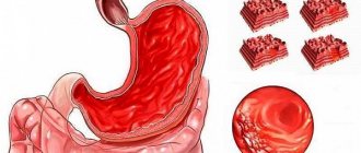
To diagnose lesions of the digestive and respiratory systems, endoscopic examinations are prescribed.
Symptoms
The clinical picture of Kaposi's sarcoma is quite varied and depends on the duration of the disease.
In the initial stages, the symptoms of sarcoma appear as reddish-bluish spots of varying shapes and sizes, as well as pink nodule-like elements, which then acquire a bluish color. As Kaposi's sarcoma progresses, the rash takes on the appearance of nodular infiltrated elements of a reddish-bluish color of various sizes. These nodules tend to merge, which leads to the formation of large lumpy foci with sharply painful ulcers. In the area of the outbreak, the skin is thickened, swollen, and purple-bluish in color. The lesions are mainly localized on the skin of the distal extremities (in 94% on the lower extremities - the anterolateral surfaces of the legs and feet) and tend to be located near the superficial veins. Often there is symmetry of the damage to the extremities.
According to its course, the tumor can be subacute, acute and chronic. The acute course of Kaposi's sarcoma is characterized by rapidly progressive symptoms and is manifested by generalized skin lesions in the form of many nodular formations on the trunk, face and limbs, as well as fever. These symptoms are accompanied by damage to internal organs and/or lymph nodes. The duration of the acute form is from two months to two years.
In the subacute course of sarcoma, generalization of skin rashes is observed much less frequently. The chronic course is characterized by gradual progression of skin rashes in the form of plaques and macular-nodular elements. The duration of the chronic form is eight years or more.
License
Kaposi's sarcoma
Kaposi's sarcoma (Kaposi's sarcoma) is a multifocal disease of a tumor nature, developing from the endothelium of blood and lymphatic vessels with possible damage not only to the skin, but also to internal organs and lymph nodes.
Etiology and key links of pathogenesis
Recently, the DNA sequence of a new herpes virus has been isolated for all forms of KS. Conventionally, this virus was named “Kaposi's sarcoma-associated herpes virus” (KSVH). It has been established that HCVH is a herpes virus type 8 (Human herpes virus type 8). Herpes virus type 8 in English literature received a second name - KSHV (Kaposi sarcoma herpes virus). The name can be translated into Russian as “herpes virus associated with KS.” Short name: HHV-8, HHV-8, KSHV, BACK. Recently, different types and subtypes of this virus have been discovered.
Taking into account modern data on the etiopathogenesis of tumor diseases, SCVG may well cause mutations in the genome region related to the control of the cell cycle, in particular the proliferation of vascular endothelial cells. The presence of hyperproliferation of transformed vascular endothelial cells, called spindle cells, does not exclude the development of events in the formations of CVH. The assumption that spindle cells are transformed endothelial cells was confirmed by the results of ultrastructural and immunohistochemical studies with markers CD31 and CD34. Tumor angiogenesis in KS was confirmed by an increased content of proteins Flk/KDR, VEGF (vascular endothelial growth factor), phosphorylated Akt enzyme, and phosphorylated p70S6 kinases involved in signaling reactions with their translocation into cell nuclei in biopsy specimens of sarcoma formations.
The likelihood of detecting SCVH increases with immunosuppression, which is manifested by a correlation with a decrease in the number of circulating CD4+ lymphocytes.
The role of SCVH in the development of KS is confirmed by changes in the production of virus defense factors called interferons in patients. Interferons are small proteins that are produced by cells already infected with the virus. Natural inducers of interferon synthesis can be the genomic RNA of viruses or the transcription product of DNA containing viruses. Interferons induce in the cell the biosynthesis of special enzymes that damage the replication cycle of viruses.
Infiltration of lesions by lymphocytes in patients with KS indicates the existence of antigens, quite possibly of viral origin, that were recognized by the immune system, resulting in the migration of lymphocytes into the skin, which is evidence of the participation of the immune system in the development of the disease.
Molecular biological studies have shown that during an experimental study of neoangiogenesis characteristic of Kaposi's sarcoma, cytokines were isolated in tumor cell cultures that stimulate the growth of cell cultures of this tumor, such as interleukin 6 (IL-6), fibroblast growth factor (3FGF), transforming growth factor p(TGFp).
Of great importance in patients with KS associated with HIV infection is oncostatin M, a cytokine produced by macrophages and activated T-lymphocytes. It is primarily produced in spindle cells and is an autocrine growth factor for AIDS-associated KS. Neutralization of oncostatin M with specific oligonucleotides reduces the growth of MC cell lines and reduces the production of IL-6. The mechanism of action of oncostatin M is mediated through tyrosine kinases, and its effect can be neutralized by the tyrosine kinase inhibitor genistein. Although HIV is not considered an etiological factor for KS, the regulatory gene product tat, which is of HIV origin, can induce the development of similar KS lesions in mice. Tat stimulates in vitro growth of spindle cells from vascular cell precursors.
In a study of oncogenes associated with KS, spindle cells were shown to overexpress the ras oncogene, in which point mutations have been identified. Int-2, an oncogenic product also known as FGF3, is expressed in 55% of KS samples.
Multicentric development, the slow evolution of the idiopathic type of KS, the possibility of regression of lesions, as well as the presence of histological signs of inflammation in the absence of symptoms of cellular atypia suggest that, at least at the beginning of its development, KS is most likely a reactive process rather than a true sarcoma.
Classification
There are 4 clinical types of KS.
- The classic or idiopathic type occurs predominantly in elderly men over 60 years of age. Favorite localization is the lower extremities. The disease progresses slowly.
- The epidemic, or African, type can account for up to 10-12% of all malignant tumors. The highest incidence is observed in young men over 20 years of age. Diseases in children are common.
- Is it an epidemic HIV-associated type? The risk of developing the disease in HIV-infected people is 20,000 times higher than in the general population. The disease progresses rapidly and affects the lymph nodes and many internal organs.
- The immunosuppressive type of KS develops both in recipients of internal organ transplants and in patients with severe diseases receiving immunosuppressive therapy. The ratio of men to women in this type of KS is 2:1, while in idiopathic (classical) it is 17:1. Currently, the immunosuppressive type of KS develops more often in recipients of internal organ transplants, in particular in 3.5% of transplant recipients. In order to prevent transplant rejection, prednisolone, azathioprine and their combinations are used, which leads to an increase in the risk of developing KS by 150-1000 times compared to the general population.
Depending on the severity of the course, the classic type of KS is divided into acute, subacute, and chronic forms.
Clinical picture
The manifestations of KS on the skin are highly variable and can be patchy, nodular and/or tumor-like.
Patients often have infiltrative plaques of round, oval or polycyclic shape, reaching the size of a palm or more. Unlike foci
nodular or spotty in nature, the surface of which is usually smooth; papillomatous growths can develop on the surface of the plaques.
The resulting tumors are usually hemispherical in shape, from 0.5 to 2 or more centimeters in diameter, and clearly rise above the surrounding apparently healthy skin.
One of the important and common manifestations of the disease are dense cells, which in most cases are localized on the lower extremities and are detected, as a rule, after the appearance of spots or nodule-like elements. However, sometimes edema precedes the appearance of these elements and for some time is the only symptom of KS.
The color of the rash varies from reddish-bluish to dark brown. The lesions have clear boundaries and are painless on palpation. Itching is also not characteristic of this disease.
Depending on the severity of the disease, the disease can last from several months to several decades.
The acute form of KS is characterized by a rapid onset, rapid generalization of the process, steady progression and death during the first year of the disease.
In the subacute form of KS, clinical manifestations are less pronounced, the process progresses somewhat more slowly and, in the absence of adequate therapy, ends in death on average after 3 years. With a timely diagnosis and rational treatment, it is possible to achieve transformation of the subacute form into a chronic one with adequate therapy. In acute and subacute forms of KS, along with spotty, nodular, infiltrative lesions, tumor diseases of various sizes are formed in large numbers. Tumors tend to ulcerate relatively quickly. As a result of the disintegration of tumors, deep ulcers of irregular shape appear with everted edges of a bluish-purple color and a lumpy, bloody-gangrenous bottom. Ulcerative detritus is absorbed, causing intoxication of the body and an increase in body temperature. Ulcers are characterized by sharp, excruciating pain. Acute and subacute forms of KS are accompanied by involvement of mucous membranes, lymph nodes, internal organs, and sometimes underlying bones. Typically, damage to internal organs occurs with minor symptoms, and more often asymptomatic.
The chronic form of KS is characterized by slow progression, limited lesions and a long course - on average 10 years, sometimes up to 15-20 years or more. Currently, the clinical picture of classical KS is undergoing certain changes. Some pathomorphism of the disease is observed. The first signs of the disease in more than one third of patients are registered in people under 50 years of age. Variants with atypical localization of the initial lesions and an unusual nature of the lesion are becoming increasingly common. In more than 10% of patients, the primary manifestations of the disease are swelling, cases of simultaneous damage to the skin and mucous membranes, and asymmetrical lesions. The nature of the course of the chronic form of classical KS changes; suddenly it acquires the features of an aggressive course characteristic of the subacute form of KS, with the development of severe edema, multiple infiltrative plaque and tumor elements, and damage to the mucous membranes. Atypical manifestations of Kaposi's sarcoma (bullous, hypertrophic) are recorded.
The most common clinical variant of endemic KS in Africa occurs in patients over 20 years of age (average age 35 years), and is 10-15 times more common in men, and is manifested by nodular, infiltrative and tumor formations, located mainly on the extremities; lymph nodes are rarely affected. This option is “locally aggressive”, with slow involvement of internal organs in the pathological process. It is characterized by high sensitivity of rashes to radiation and chemotherapy, although relapses occur more quickly than with classical KS. The lymphadenopathic variant occurs predominantly in African children aged 10 years and younger. This type is characterized by a malignant course with pronounced polyadenopathy and rapid involvement of internal organs in the pathological process, rare skin manifestations, and an unfavorable outcome within 2 to 5 years.
Epidemic KS is a unique marker of AIDS; the incidence of tumor development in this form is more than 30%. Skin lesions associated with HIV infection differ in localization from classical KS. There is a tendency to damage the scalp, proximal areas of the upper extremities, upper torso, mucous membranes, and anogenital area. The lower extremities are rarely affected. In 10-15% of cases, this type of KS occurs only with damage to the lymph nodes; in 5% of patients, internal organs are affected in the absence of changes in the skin and lymph nodes. The prognosis for life in patients with epidemic KS is unfavorable.
The immunosuppressive type of KS differs from the chronic classical form of the disease in a more aggressive course: sudden onset of the disease, the appearance of limited or multiple spot-nodule-like elements that quickly turn into tumors, frequent damage to internal organs, an extremely malignant, rapidly progressive course of the process. All these signs bring this type of disease closer to the acute and subacute forms of classical KS.
This aggressive course of the immunosuppressive type of KS is usually due to immunosuppressive therapy carried out by such patients for concomitant diseases, for example, after the start of transplantation of various organs. The first signs of KS develop in this category of patients 2-4 or more years after the start of treatment with immunosuppressive drugs. Spotty-nodule-like elements can exist for a number of years without forming a tumor; mucous membranes and internal organs are not always affected. Death, which occurs several years after diagnosis, is often associated with complications of the underlying disease for which immunosuppressive drugs were prescribed. The pathological process in those patients whose first rashes appeared on the dorsum of the feet is more benign.
SC has unusual properties:
- As a rule, it occurs in males, which is not typical for other tumors;
- The tumor initially appears in the form of multiple foci, which are symmetrically distributed in a “stocking” (“glove”) pattern.
KS never initially occurs as a main tumor with subsequent metastasis; it has a multifocal nature. The spread of foci characteristic of KS differs from metastasis in melanoma, carcinomas, and other malignant tumors, in which single or multiple metastases develop in the skin extremely rarely. True sarcomas, including rare angiosarcomas formed from vascular tissue, appear as single localized tumors that metastasize through the lymphohematogenous route to the visceral organs, most often to the lungs and liver.
Lesions of visceral organs in KS differ histologically from the microscopic signs of metastases in malignant sarcomas and carcinomas. In the affected lymph nodes, sinusoidal and capsular tumor foci are detected in combination with lymphoid hyperplasia, which is not typical for metastases to the lymph nodes in malignant tumors. Lung lesions in KS are formations on the vessels supplying the bronchi, which compress rather than invade the lung tissue, in contrast to metastases in other sarcomas. Lesions of the digestive tract in KS are characterized by involvement of the mucous membrane and submucosa in the process, in contrast to most metastatic lesions, which are intramural.
Thus, KS is an unusual tumor that has features that make it possible to distinguish it from human neoplastic diseases. On the one hand, this tumor has characteristics that distinguish it from true sarcomas (long course, multiple rashes, absence of metastases and invasive growth of tumor elements, the possibility of spontaneous involution). On the other hand, it is possible that the disease may progress to true neoplasia, when rapid generalization of the process leads to destruction of surrounding tissues, heterotopic growth, damage to visceral organs (liver, lungs, esophagus, stomach, urinary system, etc.), the appearance of symptoms of intoxication, decompensation of vital organs ending in death. The possibility of such a course of the disease brings KS closer to malignant tumors, despite the fact that in some cases the disease actually proceeds slowly, without significant destructive and decompensatory changes in the body.
Prevention
Patients with KS require constant medical supervision. They need to adhere to a work regime; work that involves standing for long periods of time, which can impair blood circulation in the extremities, is contraindicated. In severe cases of the process, a tendency towards dissemination of lesions, and resistance to therapy, patients must be referred for a medical and social examination to determine the disability group.
Epidemic type
The appearance of this neoplasm is one of the main symptoms of HIV infection. This type of Kaposi's sarcoma is characterized by:
- onset at a young age (before 40 years);
- brightly colored neoplasms;
- characterized by the obligatory involvement of mucous membranes in the process;
- unusual location of sarcoma elements: on the tip of the nose, in the oral cavity on the hard palate, upper extremities.
This type of disease is characterized by a rapid and malignant course involving lymph nodes and internal organs.
Kaposi's disease: stages of the disease
Kaposi's sarcoma is divided into several typical stages.
- Spotted. The initial degree, manifested by red spots with a bluish or dark brown (brown) tint and a smooth surface. Their diameter varies from 1 to 5 cm; individual elements can merge into large spots. The main location is the hands and feet. Subjectively, they may not manifest themselves at all or have scant symptoms (minor itching, tingling, etc.).
- Papular. It is characterized by the appearance of so-called papules - nodules, ranging in size from 2 to 10 mm, which have the shape of a sphere or hemisphere. They are located singly or grouped into clusters shaped like arcs or rings. Color - from pink to red with a brown-brown tint. Also, occasionally, a plaque with a bumpy or smooth surface may form from the spot. Horny or papillomatous growths may be observed on the surface.
- Tumor - a severe stage, accompanied by the appearance of tumor nodes. They are soft or dense, up to 50 mm, at the edges they can have bluish or brown tints. Quantity - from single to massive distribution, up to hundreds. They can merge into large lumpy neoplasms. The surface may become covered with ulcers or foul-smelling discharge. Over time, it can penetrate deep into the tissues, right down to the bones, which also affects.
Diagnostics
Diagnosis of Kaposi's sarcomatosis is carried out by an infectious disease specialist, dermatologist and oncologist. First, doctors listen to the patient and collect anamnesis, and then:
- Check for signs of disease.
- They do a biopsy.
- A histological examination is carried out to detect the proliferation of fibroblasts.
- Conduct immunological studies.
- Blood is taken for HIV testing.
The patient is also prescribed additional tests, for example, ultrasound, radiography, gastroscopy, CT scan of the kidneys, MRI of the adrenal glands and others to identify internal lesions.
Complications
The occurrence of complications of Kaposi's sarcoma depends on the stage of development of the disease and the location of the tumors. The following complications may occur:
- lymphatic edema, elephantiasis due to compression of the lymph nodes;
- bacterial infection of damaged tumors;
- limitation of motor activity of the limbs and their deformation;
- bleeding from disintegrating tumors;
- intoxication of the body caused by the breakdown of tumors;
- disruption of the functioning of internal organs when tumors are localized on them.
Some complications lead to life-threatening conditions.
Treatment of Kaposi's sarcoma
Treatment of single lesions is carried out surgically (excision of the lesion) followed by radiation therapy. Such treatment for classical Kaposi's sarcoma leads to a successful result (long-term remission) in 30-40% of patients.
If the patient has generalized Kaposi's sarcoma, and in particular, an HIV-infected person, then a complex of antiretroviral therapy, chemotherapy, interferon therapy, and radiation therapy is indicated (however, in the AIDS stage this often does not lead to the desired result).
1) Highly active antiretroviral therapy (HAART)
- the duration of such therapy should be at least a year;
- helps suppress the viral load and increase the immune status during HIV infection;
- Antiretroviral drugs can completely suppress the vital activity of one of the herpes viruses, which causes cancer - Kaposi's sarcoma.
2) Chemotherapy, for which prospidine (domestic drug), vincristine and vinblastine (rosevin), etoposide, taxol, doxirubicin, bleomycin and others are used. The drugs have pronounced side effects on the hematopoietic organs and others, which often requires the prescription of hormonal therapy (prednisolone, dexamethasone).
3) Interferon therapy.
- Purpose: interferon preparations are prescribed as an immunomodulatory effect, namely: recombinant alpha interferon 2a and 2b (intron, roferon, reaferon) or native (wellferon) in doses of 5-10 million IU/day intramuscularly, subcutaneously in long courses .
4) Local therapy includes: radiation therapy, cryotherapy, application of special gels (panretin), local chemotherapy.
Drugs that would be sufficiently effective in treating infection caused by HHV-8 have not yet been found.
Causes and risk factors
It is not possible to accurately determine the causes of the disease at this time, however, there are clear patterns that increase the likelihood of developing the disease, such as:
- Human herpesvirus type 8 - There is now some evidence that this viral disease is the main cause of sarcoma.
- Impaired immune status due to damage to the body by HIV infection.
- Some malignant tumors increase the likelihood of Kaposi's sarcoma (myeloma, Hodgkin's and others).
- Decreased immune status during the treatment of a number of diseases with drugs that have an immunosuppressive effect. These are autoimmune diseases, chemoradiotherapy for tumors.
There is an increased risk of developing the disease in men hailing from areas of central Africa and Mediterranean countries.
Prognosis of Kaposi's sarcoma
The prognosis of the disease with Kaposi's sarcoma depends on the nature of its course and is closely related to the state of the patient's immune system.
With higher levels of immunity, the manifestations of the disease can be reversible; systemic treatment gives a good effect and allows one to achieve remission in 50-70% of patients. Thus, in patients with Kaposi's sarcoma with a CD4 lymphocyte count of more than 400 μl-1, the frequency of remissions against the background of immune therapy exceeds 45%, and with a CD4 count of less than 200 μl-1, only 7% of patients can achieve remission.
Prevention
The main measure to prevent the disease is timely and correct treatment of immunodeficiency conditions. Thus, the use of antiretroviral drugs in HIV-infected patients allows for a long time to maintain the normal function of the immune system, thereby preventing the appearance of Kaposi's sarcoma.
After treatment of Kaposi's sarcoma, a thorough examination of the skin and mucous membranes is necessary at least once every 3 months, assessment of the condition of the lungs and gastrointestinal tract - at least once every six months or a year. These measures will help to detect relapse of the disease in time.
General information
Kaposi's sarcoma is a hemorrhagic multiple sarcomatosis , which is caused by multiple malignant neoplasms in the dermis of the skin and soft tissues. The pathology is named after the Hungarian dermatologist Moritz Kaposi, who first described this type of angiosarcoma .
Angiosarcomas are essentially tumors originating from the vascular perithelium and endothelial cells. They are considered extremely malignant and often metastasizing neoplasms. Multiple formations are usually localized on the skin (in 92% of cases), mucous membranes, as well as the larynx, lungs, digestive tract (in 45% of patients) and lymph nodes.
Most often occurs in people 40-50 years old, the ratio of men to women is 3:1. Despite the fact that Kaposi's sarcoma is a rather rare disease, in HIV it is the leader in the frequency of malignant neoplasms and reaches a frequency of 40-60%.