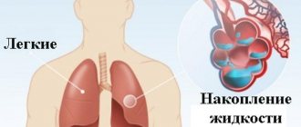
What is sarcoidosis? Sarcoidosis of the lungs.
Sarcoidosis is an inflammatory disease that can affect various human organs, but sarcoidosis mainly affects the lungs and lymph glands. In people with sarcoidosis, lumps or nodules of inflamed tissue called granulomas form in the affected organs. These granulomas can alter the normal structure or even function of the affected organ.
The exact cause of sarcoidosis is still not known. Sarcoidosis is one of the autoimmune diseases associated with an abnormal immune response, but what triggers it is not yet answered. How sarcoidosis spreads from one organ or part of the body to another is also still being studied.
Symptoms of sarcoidosis may vary depending on which organ is affected. Most patients primarily complain of a persistent dry cough, fatigue and shortness of breath. Other symptoms of sarcoidosis may include:
- The appearance of reddish bumps or spots on the skin;
- Teariness and redness of the eyes;
- Swelling and pain in the joints;
- Enlarged lymph nodes in the neck, armpits and groin;
- Nasal congestion and hoarseness of voice;
- Pain in the arms, legs and other parts of the body due to the formation of bone cysts;
Sarcoidosis can be accompanied by pathological processes in various organs and tissues of the body. For example, enlargement of the liver, formation of kidney stones, development of arrhythmia, pericarditis, and heart failure are possible. The nervous system may be affected - hearing deteriorates, convulsions, mental disorders (dementia, depression, psychosis), meningitis appear.
In some people, symptoms of sarcoidosis appear suddenly and disappear within a short time. Others may not notice any changes at all, although the organs are affected. Or it may be that the symptoms appear slowly and gradually, and recur periodically.
Sarcoidosis most often affects people between the ages of 20 and 40. Moreover, women are diagnosed with this condition more often than men.
A must read! Help with hospitalization and treatment!
The incidence of sarcoidosis in Belarus is 3.9 cases per 100 thousand people. The main contingent of patients with this pathology are young people (the peak is 25–35 years old) who lead an active lifestyle. Women get sick more often than men.
Natalia Glutkina, senior lecturer of the 1st Department of Internal Diseases of the Grodno State Medical University, Candidate of Medical Sciences. Sciences. Etiology and symptoms
Sarcoidosis (Besnier-Beck-Schaumann disease) is an inflammatory disease of unknown etiology, which is characterized by multisystem damage to various organs (usually lung tissue and intrathoracic lymph nodes) and the formation of epithelioid granulomas in the affected tissues. Since the majority of patients are people of working age, the treatment of this disease has an important social aspect.
Currently, there is no consensus on the etiology of this disease. There are several hypotheses related to infectious agents acting as a trigger (mycobacteria, Chlamydophila pneumoniae and others), the environment, smoking, and heredity.
There are several options for the course of the disease : spontaneous regression, regression during therapy, progression with the development of respiratory failure and/or severe extrapulmonary damage, as well as an undulating course, relapse (according to various studies, the threat ranges from 8% to 74%). The absence of spontaneous remission within 2 years indicates a chronic or persistent course of the disease.
In the early stages, a low-symptomatic clinical picture predominates (in 30–70% of cases), detected only by the presence of characteristic changes on a chest x-ray. In some patients, the onset may be acute, similar to Löfgren's syndrome (bilateral enlargement of the lymph nodes of the roots of the lungs according to radiography, erythema nodosum, fever, myalgia, weight loss and joint pain).
Extrapulmonary symptoms are caused by the presence of non-caseating granulomas in various organs and tissues: skin lesions (the appearance of erythema nodosum, mainly localized on the anterior surface of the lower extremities ( see Fig. 1 )); lupus pernio (induration and bluish-red coloration of the lips, cheeks and nose, possible deformation of the cartilage with a change in the shape of the nose); lesions of the eye structure (usually anterior or posterior uveitis), the cardiovascular system (various types of arrhythmias, conduction disorders and chronic heart failure). Changes in the nervous system occur in 10% of cases (facial nerve palsy, peripheral neuropathies and neuromuscular lesions).
One of the manifestations of damage to the hematopoietic system may be fever of unknown origin in combination with lymphopenia. Involvement of the liver in the pathological process can manifest itself as an asymptomatic increase in the level of liver enzymes, less often - hepatomegaly; kidneys - subclinical proteinuria, tubulointerstitial disorders. In less than 6% of patients, this disease manifests as Heerfordt's syndrome (unilateral or bilateral mumps, fever, enlarged parotid glands, anterior uveitis and facial paralysis).
Rice. 1. Patient with stage 2 Beck’s sarcoidosis. - pulmonary-mediastinal form (in the area of both legs, hyperpigmentation of the skin is determined in places preceding erythema nodosum).
Diagnostics
Laboratory data
The acute course of sarcoidosis is characterized by accelerated ESR, less commonly monocytosis, and lymphopenia. In a biochemical study, an increase in fibrinogen, beta-lipoproteins, serotonin, C-reactive protein, and sialic acids may be observed. Sometimes with sarcoidosis there is an increase in serum calcium and calciuria, but more often hypercalcemia is not clinically manifested and is transient.
Bronchopulmonary examination data
With sarcoidosis of the intrathoracic lymph nodes (HTLU), there is an expansion of the spurs of the trachea and bronchi, narrowing and deformation of the lumens of the bronchi, “sarcoidosis ectasia” - dilated, thickened, tortuous vessels in the form of a sparse network or individual large plexuses. In sarcoidosis of the upper lymph nodes and lungs, such changes are less pronounced, but sarcoidosis itself, associated with granulomatous lesions of the bronchial tree, is more common. With pulmonary sarcoidosis, inflammatory processes in the bronchial tree come to the fore. Involvement of the intrathoracic lymph nodes occurs in more than 90% of cases ( see Fig. 2 ). Asymmetrical increase in VGLU is observed in 10% of cases.
Rice. 2. X-ray of the chest organs in stage 1 sarcoidosis. (the pathology was detected during a routine preventive examination, asymptomatic).
There are 5 stages of sarcoidosis of the chest organs, however, the concept of stages is quite arbitrary, and the transition of the disease from one stage to another is rare ( see table ). The key for correct diagnosis is performing computed tomography of the chest organs to assess the volume of the lesion and histological verification . High-resolution CT helps to evaluate changes around the bronchi, and in the interstitium - lesions located along the vascular-bronchial cords and under the pleura. Small lesions may be accompanied by “frosted glass”. In 1/3 of patients, atypical changes are detected: large shadows (2–4 cm in diameter) or nodular changes with the presence of enlarged lymph nodes ( see Fig. 3 ).
Rice. 3. CT picture of interstitial nodular changes in the lungs, enlargement of the intrathoracic lymph nodes, characteristic of sarcoidosis. For neurosarcoidosis, an informative diagnostic method is MRI (hydrocephalus, enlargement of the basal cisterns, single or multiple granulomas are detected), this method is especially relevant for damage to the meninges . However, in assessing the condition of the lung tissue it is inferior to computed tomography. Positron emission tomography allows you to obtain reliable information about the activity of the process.
Pulmonary sarcoidosis requires differential diagnosis with a large number of pulmonary diseases, which are based on morphological verification of the diagnosis.
Histological verification of sarcoidosis is based on identifying its main histological structure - compact non-caseating epithelial cell granuloma. In the center of the granuloma, granular masses stained with eosin may be visible. This zone resembles fibrinoid necrosis, but is not a zone of caseous necrosis. In the center of individual granulomas, giant Pirogov-Langhans cells can be seen, and cells of foreign bodies are also found. In the cytoplasm of these cells, crystalloid inclusions, asteroid bodies and Schaumann bodies are often found, which, however, are not specific for sarcoidosis.
Treatment and prognosis
The main goal of treatment for sarcoidosis is to reduce symptoms and improve the quality of life of patients. But before starting treatment, it is necessary to compare the appropriateness of the prescription with the risk of developing side effects from the use of corticosteroid, cytostatic and biological therapy.
All patients with sarcoidosis are advised to limit hyperinsolation, dairy products and other foods high in calcium. There is no etiotropic therapy for sarcoidosis.
For asymptomatic patients, treatment is not indicated (possible side effects that develop when using glucocorticosteroids have a greater impact on the quality of life of patients than the manifestations of the disease itself); it is possible to use pentoxifylline at a dose of 600–1200 mg per day, alpha-tocopherol 200 mcg 2 times per day (at least 6 months) in this category of patients.
Systemic glucocorticosteroids should be used in patients with a progressive course of the disease according to X-ray and functional breathing studies, in the presence of extrapulmonary manifestations that impair organ function, or the development of lupus pernio.
For pulmonary sarcoidosis, the initial dose of prednisolone is 25–30 mg per day for 4 weeks, with a possible subsequent gradual reduction in the dose to a maintenance dose of 10 mg to control symptoms and disease progression (continued for 12–24 months). After 3 months from the start, it is necessary to evaluate the effect of glucocorticosteroid therapy.
Pulse therapy with glucocorticosteroids, as well as therapy with 4-aminoquinoline drugs, is carried out in exceptional cases (in case of multiple organ damage, refractoriness to usual doses of therapy).
If glucocorticoids are ineffective or undesirable effects occur, alternative therapy with immunosuppressants is possible : methotrexate, azathioprine, cyclosporine and cyclophosphamide.
In severe forms of sarcoidosis or in case of ineffectiveness of the above treatment options, it is possible to use TNF-α antagonists (infliximab has the greatest evidence base).
To increase the sensitivity of the effects of pharmacological drugs, it is possible to use an extracorporeal method - plasmapheresis , by improving microcirculation, leading to the unblocking of cellular receptors and stabilization of cell membranes.
The variety of clinical symptoms of sarcoidosis significantly lengthens the process of diagnosis and leads to a large number of diagnostic errors (up to 70%), therefore the search for new diagnostic methods, timely and adequate treatment of this pathology are extremely important for the prevention of complications, the formation of which largely depends on the timing of diagnosis.
2. Diagnosis of sarcoidosis
There are various ways to diagnose sarcoidosis, and usually a comprehensive examination is necessary for an accurate diagnosis, since all symptoms and laboratory results can be characteristic of other diseases. Therefore, if you suspect sarcoidosis, it is very important to find a good doctor who will carefully study the clinical picture of the disease, symptoms and medical history. In general, the following can be used to diagnose sarcoidosis:
- X-ray of the chest, which allows you to detect infiltrates in the lungs with sarcoidosis of the lungs or tumors of the lymph nodes;
- Computed tomography allows for an even more thorough examination of the lungs and lymph nodes than a chest x-ray;
- Lung tests (respiratory function tests) can show how well your lungs are working;
- Bronchoscopy is performed to examine the bronchi, perform a biopsy (obtain a small tissue sample for further examination), and detect granulomas.
- Bronchoscopy is performed using a bronchoscope (a small flexible tube) that is passed into the trachea (windpipe) and into the bronchi.
Visit our Pulmonology page
What is inflammation of the lymph of the lung
Lymphatic small and large vessels of the lungs perform the function of absorbing and removing protein fluid, returning it to the blood circulation. Various substances and microorganisms in the liquid pass through a biological filter and can cause various reactions in the lymph node. When pneumonia occurs, the vascular and lymphatic systems and mediastinal lymph nodes are involved in the inflammatory process. Pneumonia is characterized by an inflammatory process not only in regional lymph nodes; the pathological process can also involve extrathoracic and distant lymph nodes.
The inflammatory process spreads to the lymph nodes along the bronchi and trachea. It is not always possible to see the enlargement of certain groups of lymph nodes due to their location. An X-ray examination may not show an increase in hilus, bronchopulmonary nodes, which are often overlapped by the branches of the pulmonary artery or the shadow of the heart; to determine the pathological state of the bifurcation lymph nodes, an x-ray examination is carried out in a lateral projection. To determine the inflammatory process in the lymph nodes of the lungs, X-ray examination is carried out in oblique, sagittal and lateral projections.
3. Treatment of the disease
There are no targeted treatments for sarcoidosis. However, the patient's condition may improve on its own over time. Many people with sarcoidosis have mild symptoms of the disease, for which no treatment is required. In other cases, if necessary, therapy is given to reduce the severity of sarcoidosis symptoms and maintain normal functioning of the affected organs.
Treatment for sarcoidosis is usually carried out in two ways - general health measures (regular medical examinations, smoking cessation, healthy eating, weight control and other healthy lifestyle principles) and drug treatment. Drug treatment for sarcoidosis is used to relieve symptoms and reduce inflammation of affected tissues.
What if the disease progresses?
For many people with sarcoidosis, the disease appears for a short time and then disappears without a trace. A person may not even know that he was sick. Between 20 and 30% of people suffer permanent lung damage as a result of pulmonary sarcoidosis. In approximately 10-15%, pulmonary sarcoidosis takes a chronic form. In some cases, the disease can lead to dysfunction of the affected organ. Sarcoidosis is rarely fatal and usually results from complications in the lungs, heart, or brain.
About our clinic Chistye Prudy metro station Medintercom page!
Why is inflammation of the lymph nodes in the lungs dangerous?
Lymph nodes play an important role in pneumonia. During the inflammatory process, a reaction of the lymph node to pathogenic microorganisms occurs - as a result of spasm and closure of the lymphatic vessel, inflammatory edema begins to develop. This reaction stops pathogenic microorganisms at the site of the inflammatory process and prevents the penetration of microbes into the bloodstream of the circulatory system. With pneumonia, the development of perilymphatic foci is observed, which are located along the lymph nodes.
Such changes are also observed in lymphogenous carcinomatosis (tumor cells spread through the lymphatic vessels) and sarcoidosis (a systemic disease characterized by damage to organs and systems of the body, damage to the lymph nodes). The lymphatic vessel may close, the drainage and cleansing function of the lymph nodes may be disrupted, and the infectious process will progress. Enlargement and pain of the lymph nodes of the chest cavity are observed with tuberculosis; enlargement of cervical, intra-abdominal, intrathoracic, axillary lymph nodes is typical for patients with HIV infection.
Possible reasons
To date, there is no evidence that any of the following causes sarcoidosis. They are described at the level of hypotheses.
Infectious factors
. According to scientists, the triggers of the disease include:
- the causative agent of Lyme disease (Borrelia burgdorferi);
- microorganism Chlamydophila pneumoniae;
- mycobacteria;
- certain types of viruses (for example, hepatitis C, herpes);
- bacteria Propionibacterium acnes.
Environmental factors
. It is assumed that contact with smoke, agricultural dust and mold can provoke granulomatous changes in the internal organs.
Heredity
. Some scientists believe that relatives of patients with sarcoidosis have an increased risk of developing this disease. In this case, specific genetic causes are identified (polymorphism of certain genes).
What are the symptoms of the disease?
Most often, the onset of the disease is asymptomatic, the disease does not manifest itself in any way, so the most important thing that can be advised is to regularly undergo preventive X-ray examinations, which allows you to detect not only tuberculosis, oncological diseases, but also, not least, sarcoidosis. I would especially like to emphasize the importance of not skipping preventive X-ray examinations for women after childbirth and cessation of breastfeeding. Sometimes sarcoidosis can begin acutely, with a rise in temperature and the appearance of red-purple painful spots on the legs, accompanied by pain in the joints. In such cases, it is also necessary to take an x-ray, which may reveal enlarged intrathoracic lymph nodes. This is the so-called Löfgren's syndrome, which is very characteristic of sarcoidosis. It is very likely that young Mozart suffered from sarcoidosis, since his father’s letters contain a description of the typical picture of this syndrome.
Symptoms
The main manifestations of sarcoidosis are constant fatigue, fatigue, and decreased ability to work. However, there is no direct relationship between the severity of these symptoms and the extent of damage to the liver or other organ. After the disappearance of granulomatous inflammation, general weakness usually disappears. A characteristic set of symptoms can also be observed during the acute onset of the disease, namely:
- Löfgren's syndrome (joint syndrome, fever, erythema nodosum, hilar lymphadenopathy);
- Heerfordt-Waldenström syndrome (mumps, uveitis, fever).
Principles of treatment
Diagnosis of sarcoidosis is carried out by a physician. At an early stage of the disease, in the absence of obvious symptoms, treatment is not carried out. This is due to the fact that sarcoidosis can go away on its own in 3-6 months. However, throughout this period the patient’s condition must be monitored.
If there are obvious signs of the disease, the basis of treatment is hormonal therapy. Hormones are initially prescribed in high doses, then switched to maintenance. Sarcoidosis is prone to relapse, so hormone therapy may be repeated several times.
The prognosis for the course of the disease is generally favorable. But some patients experience severe dysfunction of vital systems, which leads to disability.
Stages of sarcoidosis
Experts classify the stages of sarcoidosis in different ways. Some adhere to three stages of pathology development. Others use a more detailed classification and distinguish five stages. We will focus on the last option and describe the five stages of pathology development. It is this classification that is included in international recommendations for the treatment of the disease.
0 – At this stage, manifestations of the disease and changes on a chest x-ray may not be observed. There is no involvement of the lungs, but this does not exclude sarcoidosis of other organs.
1 – During this period of development, the disease may manifest itself with minor symptoms: cough, shortness of breath on exertion, weakness and low fever. The intrathoracic lymph nodes are enlarged. The lung parenchyma, that is, the tissue of the respiratory organ, is not changed.
2 – At this stage, symptoms become more obvious. These include temperature up to 40 °C, chest pain, weakness and sweating, cough. The lymph nodes are enlarged, the lung parenchyma has already begun to undergo changes.
3 – Neurological changes are added to the symptoms. The pulmonary parenchyma is affected, but the lymph nodes are not enlarged.
4 – At this stage, irreversible pulmonary fibrosis is detected. Shortness of breath progresses, cough intensifies. Due to lack of oxygen, neurological symptoms become more severe. Granulomas grow in the lungs and other organs. The risks of complications increase.
Treatment of sarcoidosis
In the absence of pronounced active organ lesions, acute sarcoidosis (Lefgren's syndrome), it is possible to prescribe only symptomatic therapy (NSAIDs) under the supervision of a physician, with regular dynamic examination.
With the development of progressive damage to the lungs, kidneys, heart, and other target organs of sarcoidosis, immunosuppressive therapy is prescribed, which includes the following classes of drugs:
- glucocorticoids (prednisolone, methylprednisolone) are the first line of therapy;
- cytostatic drugs (methotrexate, azathioprine, mycophenolate mofetil, cyclosporine, chlorambucil, cyclophosphamide) - are prescribed as steroid-sparing agents, with a long-term active course of the disease, the development of steroid dependence;
- genetically engineered immunobiological drugs from the anti-TNF group (infliximab, adalimumab) - are prescribed in case of insufficient effect or intolerance to therapy with corticosteroids and/or cytostatic drugs.
Additionally, in the presence of sarcoidosis of the lungs, skin, articular syndrome, or impaired phosphorus-calcium metabolism, patients may be prescribed drugs of the aminoquinoline series (hydroxychloroquine, chloroquine).
What is the prevention of sarcoidosis?
Prevention of sarcoidosis has not been developed, since its cause is not clear. But Immunostimulants, including those of plant origin, inducers of endogenous interferon, sets of antigens of various bacteria are potentially dangerous as triggers of sarcoidosis.
Diet – avoid large amounts of calcium and vitamin D. Do not use multivitamin complexes containing calcium.
Mode – physical training and limiting exposure to ultraviolet radiation are recommended.
The main thing I would like to advise you to do is to learn to cope with stress, enjoy life, communicate with friends and family in order not to get sarcoidosis. In many countries, for example, in Germany, after a diagnosis of sarcoidosis is made, the patient is first of all referred to a psychologist.
Symptoms of sarcoidosis
The disease can occur for a long time without specific symptoms. At the initial stage, sarcoidosis is often detected accidentally during a preventive X-ray examination of the lungs.
General nonspecific signs of pathology include:
- Weakness
. Most often it occurs in the morning, sometimes accompanied by painful sensations in the muscles and headaches. - Temperature increase
. Fever in most cases accompanies sarcoidosis, which occurs with damage to the lymph nodes and eyes. - Reducing body weight
. Weight does not decrease immediately, but several months after the onset of the disease. Weight loss occurs due to metabolic disorders, which leads to deterioration in the absorption of nutrients. - Enlarged lymph nodes
. The cervical and thoracic groups of lymph nodes are more susceptible to inflammation. On palpation, such nodes are dense, painless and mobile.
Specific symptoms of sarcoidosis depend on the location of the granulomas:
- When granulomas form in the lungs,
in 90% of cases shortness of breath, dry or wet cough, and heaviness in the chest appear. - Sarcoidosis with eye damage
leads to the spread of small granulomas to the cornea, retina, sclera, and outer membranes of the eye. As a result, lacrimation, redness of the eyes, decreased vision, sometimes to the point of complete blindness, are disturbing. - When the skin is affected,
the nodules look like rounded formations raised above the body. The surface of the skin in these places becomes reddish. Sarcoidosis of the skin can affect the lower extremities, gluteal region, and face. Sometimes erythema nodosum appears - painful red nodes that turn yellow over time. Erythema nodosum often occurs with signs of arthritis - swelling and pain in the elbow, ankle, and wrist joints; Sarcoidosis involving the nervous system can cause facial paralysis. The disease is manifested by headaches, memory loss, weakness of the muscles of the limbs, and in rare cases, seizures.
The most likely forms of sarcoidosis are described above. But there have also been cases of granulomas affecting the heart, spleen, genitourinary organs, oral cavity, and digestive organs.
Sarcoidosis occurs in waves - periods of subsidence of symptoms are replaced by another exacerbation. In some patients, self-healing occurs. Complications of the disease may include respiratory failure, severe impairment of liver and kidney function. If the heart is damaged, death can occur.
The diagnosis is made on the basis of x-ray examination, blood tests (an inflammatory reaction is detected), and histological examination of granuloma biomaterial.


