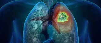
Lobar pneumonia is an acute infectious disease in which one or more lobes of the lung are affected, fibrinous effusion appears in the alveoli, and fibrinous deposits appear on the pleura. Croupous pneumonia affects mainly adults. The disease is characterized by a pronounced clinical picture and symptoms of intoxication. Patients with lobar pneumonia are admitted to the therapy clinic.
To examine patients at the Yusupov Hospital, doctors use modern equipment from leading European, American and Japanese companies. Pulmonologists use European treatment protocols and take an individual approach to the choice of treatment method for each patient. Medicines are administered through the digestive tract, intramuscularly, intravenously and by inhalation. Thanks to complex treatment, the length of stay of patients in hospital is reduced.
Signs of lobar pneumonia in adults
With lobar pneumonia, symptoms appear acutely. Key ones:
- a sharp increase in temperature to 39 - 40 ° C;
- severe chills, weakness;
- chest pain when breathing;
- cough - at first dry, but quickly becomes wet, with rust-colored sputum;
- severe shortness of breath;
- wheezing that may be heard when breathing;
- rapid heartbeat, low blood pressure;
- pale skin, nausea, muscle weakness.
The general condition is serious, the patient needs the help of doctors in the hospital.
Symptoms of lobar pneumonia
Lobar pneumonia has an acute onset. In patients in full health, body temperature rises to 39 ° C, chills and chest pain appear. In the initial stage of the disease, the cough is dry, then it becomes productive, with the release of “rusty” sputum. There is severe shortness of breath, the chest on the affected side lags behind when breathing.
In the initial phase of inflammation, percussion reveals a dull tympanic sound over the lesion. During auscultation, harsh breathing with prolonged exhalation, mild crepitus, and wet and dry rales in a limited area are heard. In the compaction phase of lobar pneumonia, the following symptoms appear:
- a sharp increase in vocal tremors, bronchophony during palpation of the chest;
- with percussion – dull sound;
- vesicular breathing is not audible, crepitus disappears, and pleural friction noise is often heard.
In the resolution phase, vocal tremors gradually normalize, bronchophony disappears, and abundant, sonorous crepitus appears over a long period of time. Loud fine-bubble rales are heard, bronchial breathing gradually gives way to harsh and then vesicular breathing.
When examining the cardiovascular system, a rapid pulse is determined. In the case of severe lobar pneumonia, it is poorly filled, arrhythmic, blood pressure is reduced, and the heart sounds are muffled.
Stages of lobar pneumonia in adults
Lobar pneumonia proceeds according to a special scenario, with a change of stages with typical changes in the lung tissue.
Stage one - high tide. A lot of blood flows to the lung tissue, which is why it turns red, and blood stagnates in small vessels. The stage lasts from 12 - 14 hours to 3 days.
Stage two – red liver. During this period, some of the fluid and red blood cells leak out of the small vessels of the lungs into the alveoli, which is why they work poorly. The affected part of the lung becomes dense, similar in appearance to liver tissue. Fibrin accumulates inside the lung sacs, a sticky substance that gives the tissue its density. This stage lasts up to 3 days.
Stage three – gray hepatization. Red blood cells stop entering the lung tissue, they are replaced by leukocytes and fibrin, epithelium. Therefore, the color of the lungs changes to gray and greenish. The stage lasts from 2 to 6 days.
Stage four – resolution. Gradually, fibrin dissolves, the alveoli are cleared, the lungs are restored and begin to breathe normally. This process is the longest, can last up to 2 - 3 weeks.
How does infection occur?
The causative agents of lobar pneumonia are pneumococci types I-IV. Less commonly, the disease is caused by Friendler's diplobacillus. As a rule, inflammation manifests itself acutely, against the background of absolute health and lack of contact with infected persons. Based on this, we can conclude that the infectious agents were previously in the upper respiratory tract, but their reproduction was restrained due to the work of the immune system. Its weakening is one of the leading factors in the development of lobar pneumonia.
From the point of view of modern medicine, lobar pneumonia is considered an infectious-allergic disease. In the vast majority of cases, the disease develops as a result of infection of the lungs with type I and II pneumococci. The polysaccharide capsule of pneumococci ensures their virulence and also provokes pronounced sensitization of the body.
Conditions that are necessary for the development of lobar pneumonia:
- Primary penetration of pneumococci into the body and development of inflammation. Moreover, its focus may not be located in the lung tissue, but may have a different localization.
- Sensitization of the body to a certain type of pneumococcus and re-entry of infection.
So, lobar pneumonia develops under the condition of secondary infection with pneumococci. An important condition is the fact of re-infection at the peak of sensitization of the body to microbes of a certain type. They can enter the lungs through blood, lymph or airborne droplets.
If all conditions are met, a violent reaction develops in the body, which is similar to what occurs when a foreign protein is introduced. A whole chain of morphological changes is launched in the lungs, which were described by Laennec (flush, red and gray hepatitis, resolution). Inflammatory exudate accumulates in the alveoli, a significant proportion of which is fibrin.
Modern methods of treatment
To treat lobar pneumonia, your doctor will prescribe two antibiotics.
One drug is administered intravenously, the second intramuscularly. Usually these are drugs from the group of penicillins and cephalosporins. Stronger medications are required less often. Additionally, complex therapy is required:
- immunocorrection (blood plasma and immunoglobulins are administered);
- correction of blood clotting disorders (intravenous blood thinning solutions, heparin);
- normalization of the protein composition of the blood (intravenous albumin, retabolil);
- oxygen (supplied through a nasal catheter or mask);
- hormonal drugs (for severe cases, prednisone is used for a short course);
- antioxidant therapy (ascorbic acid, rutin);
- bronchodilators (Berodual, Eufillin, Atrovent);
- expectorants (ACC, Bromhexine, Ambrobene, Lazolvan);
- antipyretic drugs (Nurofen, Paracetamol, Ibuklin);
- physiotherapy, massage, exercise therapy.
As the patient’s condition improves, he undergoes a course of rehabilitation to completely eliminate foci of inflammation in the lungs.
Features of the course of the disease in children
Children rarely experience fever and chills, and they do not complain of pain in the side.
An atypical course of lobar pneumonia is observed in young children. At the onset of the disease, there is no cough, but other symptoms occur: dry mouth, bloating, nausea and vomiting, abdominal pain, pale skin, rapid breathing, hyperexcitation or lethargy, enlarged liver. Sometimes there is a stiff neck, headache, convulsions, delirium and hallucinations. The combination of such symptoms can cause an incorrect diagnosis (meningitis). As pneumonia progresses, meningeal signs give way to the classic clinical picture of pneumonia.
Croupous pneumonia rarely develops in children. People aged 18-40 years are most susceptible to it.
In children 7-16 years old, symptoms do not differ from those that occur in adults. Body temperature stabilizes on days 5-9 from the onset of the disease. At the same time, inflammation in the lungs subsides.
Popular questions and answers
Pulmonologist Marina Samoilova told us why lobar pneumonia is so dangerous and how to treat it.
What complications can occur with lobar pneumonia?
If a person has a weakened immune system or treatment is started untimely or incorrectly, complications are possible in the form of:
- abscess (purulent melting of lung tissue); gangrene of the lung;
- purulent pericarditis (inflammation of the lining of the heart);
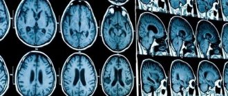
- purulent brain damage;
- blood poisoning;
- respiratory failure and even death.
Pneumococcal pneumonia, even in our time, is a fairly common cause of death, especially in older people, smokers, people suffering from alcoholism, with cancer and systemic diseases, and HIV-infected people.
Story
Pneumonia was first described by Hippocrates around 460 BC, but until the 19th century it was not known that it was an infection of the lungs and not a symptom of other diseases. The first clinical and pathological manifestations were described by the French physician René Laennec in 1819, and in 1842 the Austrian pathologist Karl Rokitansky was the first to differentiate pneumonia into lobar and bronchopneumonia. The infectious nature was discovered in 1875 by the German pathologist Edwin Klebs, who observed bacteria under a microscope, and in 1884 Karl Friedlander and Frenkel, Albert were able to identify the two most common pathogens of pneumonia at that time - Streptococcus pneumoniae (pneumococcus) and Klebsiella pneumonia. By 1929, the International List of Causes of Death had 94 terms for pneumonia; later in the ICD-10 classifier, many of the descriptive terms that included pneumonia were eventually removed, but it is still listed in descriptive terms for some infectious and noninfectious causes, including complications from certain diseases and procedures. In the 1930s, penicillin developed treatments for bacterial pneumonia, but pneumonia is still the leading cause of death among children under 5 years of age.
23.01.2021 6712
Is it possible to treat lobar pneumonia with folk remedies?
As a rule, patients with lobar pneumonia should be hospitalized for constant monitoring by medical staff and emergency measures at the slightest sign of complications.
Often such patients experience nausea, vomiting, bowel movements, and confusion, so medications and solutions are administered intravenously. Thus, we cannot talk about treatment at home without the participation of qualified specialists. Fortunately, in our time, subject to a healthy lifestyle, such serious diseases as lobar pneumonia are becoming less and less common, and with modern treatment methods, in most cases it is possible to avoid serious complications and save the patient’s life.
Published on the portal kp.ru
Prevention
Prevention of pneumonia is an important factor in reducing child mortality. Vaccination against Haemophilus influenzae type B, pneumococcus, measles and whooping cough is the most effective way to prevent pneumonia. For young children, nutrition plays an important role, particularly exclusive breastfeeding for the first 6 months of life. Reduces the incidence of illness among children and combats air pollution, for example, through the use of environmentally friendly cookstoves in residential buildings. In overcrowded homes, providing hygienic conditions can help reduce disease incidence.
In health care settings, effective measures to reduce the spread of infection include hand washing and the use of gloves and masks, and patient isolation is recommended.
Vaccinal prevention
Vaccinal prevention of pneumococcal infections
According to the position of WHO and the Russian Respiratory Society, “Vaccination is the only way to prevent the development of pneumococcal infection.” In the Russian Federation, 94% of all etiologically deciphered cases of complicated pneumococcal infection in children are caused by pneumococcal community-acquired pneumonia. Pneumococcus is the cause of up to 76% of community-acquired pneumonia in Russian adults. For vaccination against pneumococcal infection of persons over 2 years of age in the USA since 1983, and in the Russian Federation since 1999, polysaccharide polyvalent vaccines containing antigens of 23 serotypes, causing up to 90% of invasive diseases of pneumococcal etiology, have been successfully used. Vaccination is carried out once, followed by revaccination for patients from high-risk groups (over 65 and immunocompromised individuals) after 5 years. The effectiveness of polysaccharide vaccines reaches 80%, but may be lower in elderly people, patients with immunodeficiency conditions, and in children under 2 years of age. These vaccines cause the formation of T-independent B-cell immunity.
Publications in the media
Pneumonia is a group of acute infectious (mainly bacterial) diseases, different in etiology, pathogenesis and morphological characteristics, characterized by focal damage to the respiratory parts of the lungs with the obligatory presence of intra-alveolar exudation. Bacterial pneumonia is pneumonia of bacterial etiology. Classification • According to the conditions in which the disease developed •• Community-acquired pneumonia - acquired outside a medical institution (synonyms: home, outpatient) •• Nosocomial pneumonia - acquired in a medical institution (synonyms: hospital, nosocomial) •• Aspiration pneumonia •• Pneumonia in individuals with severe immune defects (congenital immunodeficiency, HIV infection, iatrogenic immunosuppression, etc.) • Along the course •• Mild - does not require hospitalization •• Severe - hospitalization is required.
Incidence • 236.2 cases per 100,000 adolescents 15–17 years of age • 522.8 cases per 100,000 population under 14 years of age • Community-acquired pneumonia - 1,200 cases per 100,000 population per year • Hospital-acquired pneumonia - 800 cases per 100,000 hospitalizations per year . The predominant age is under 20 and over 60 years. Predominant gender - no significant differences were found by gender. Etiology • Streptococcus pneumoniae - most common (30-50%) • Haemophilus influenzae (10-20%) • Atypical pathogens - Chlamidophila pneumoniae, Mycoplasma pneumoniae, Legionella pneumophila (8-25%) • Typical but rare (3-5 %) include Staphylococcus aureus, Klebsiella pneumoniae (less commonly other enterobacteria) • Moraxella catarrhalis (Branhamella catarrhalis) • Most often, community-acquired pneumonia is caused by pneumococci (their sensitivity to penicillin in many countries is significantly reduced) • In very rare cases of community-acquired pneumonia - Pseudomonas aeruginosa (with cystic fibrosis, bronchiectasis), in persons with severe immunodeficiency - Pneumocystis carinii • Escherichia coli • Anaerobic microorganisms • Atypical pneumonia.
Risk factors • Recent acute respiratory viral infection • Renal failure • Cardiovascular diseases • COPD • Immunodeficiency conditions: diabetes, chronic alcoholism, AIDS, malignant neoplasms • Dysbiosis • Risk factors for hospital-acquired pneumonia •• Mechanical ventilation •• Early postoperative period •• Dysbacteriosis • Risk factors aspiration pneumonia •• Impaired consciousness •• Convulsive seizures •• Diseases of the central nervous system •• Anesthesia •• Reflux esophagitis. Pathogenesis . Main pathogenetic mechanisms • Aspiration of oropharyngeal secretions (the main route of infection) • Inhalation of an aerosol containing microorganisms • Hematogenous spread of pathogens from an extrapulmonary source of infection (for example, with endocarditis, with septic thrombophlebitis) • Direct spread of infection from neighboring affected organs (for example, with liver abscess ) or as a result of injury and infection of the chest organs. Pathomorphology . Segmental, lobar or multifocal peribronchial compaction with stages of red (intra-alveolar exudation and erythrocyte diapedesis) and then gray (fibrous organization of intra-alveolar exudate) hepatization.
Clinical picture. • Complaints •• Cough with mucopurulent (sometimes “rusty”) sputum •• Chest pain when breathing (with concomitant pleurisy) •• Shortness of breath •• Weakness, fatigue •• Night sweats. • Intoxication syndrome •• Fever •• Tachycardia •• Tachypnea •• Hyperhidrosis •• Myalgia •• Headaches •• Anorexia. • Objective examination data •• Cyanosis •• Percussion: dullness of percussion sound caused by infiltration or pleural effusion •• Auscultation ••• Moist fine rales and/or crepitus (heard on inspiration) ••• With extensive infiltration or pleural effusion, breathing is weakened • •• Pleural friction noise with dry pleurisy •• In severe cases, meningeal signs and disturbances of consciousness (for example, disorientation and anxiety) may appear •• In 20% of cases, objective signs may be mild or absent. Laboratory tests • Leukocytosis with a shift of the leukocyte formula to the left •• Leukocytosis more than 10-12109/l is characteristic of a bacterial infection •• Values more than 25109/l or leukopenia below 3109/l indicate a poor prognosis • Hyponatremia • Increased activity transaminases • Bacteriological blood test to identify the pathogen (positive result in 20–30% of patients with community-acquired pneumonia, especially before the start of antibacterial therapy) • At the height of active inflammation and intoxication, protein may appear in the urine • Bacteriological and bacterioscopic examination of sputum: microscopy of a stained smear according to Gram, and culture of sputum obtained during deep coughing • Bacteriological examination of material obtained during bronchoalveolar lavage and thoracentesis • In the presence of pleural effusion and pleural puncture - examination of pleural fluid: counting leukocytes with a leukocyte formula, determination of pH, protein content, LDH, microscopy of a Gram-stained smear, culture for aerobic, anaerobic bacteria and mycobacteria • Study of the immune status of persons with suspected immunodeficiency. Special studies • Chest X-ray is a mandatory research method for pneumonia, allowing to visualize areas of infiltration of lung tissue (take into account the shape, size and location), assess the dynamics of the process •• The prevalence of infiltration, the presence of pleural effusion and signs of destruction of lung tissue reflect the severity of the disease and significantly influence the nature of treatment • CT scan of the lungs is performed if destruction or neoplasm is suspected • Fiberoptic bronchoscopy with microbiological and cytological examination of the biopsy specimen - if tuberculosis and tumor diseases are suspected • FVD study - for differential diagnosis with respiratory distress syndrome • With extensive infiltration, massive pleural effusion, In the presence of COPD, it is advisable to evaluate arterial blood gases, which may be the basis for hospitalization and low-flow oxygenation • The study of capillary blood gases is not very informative.
Diagnostic tactics , diagnostic algorithms. The diagnosis of pneumonia is considered definite if the patient has radiologically confirmed infiltration of the lung tissue and at least two of the following signs: • acute febrile onset of the disease (body temperature more than 38 ° C); • cough with sputum; • listening to local crepitus, shortening of percussion sound; • leukocytosis more than 10109/l and/or shift of the leukocyte formula to the left more than 10%. Differential diagnosis • Pneumonia of non-bacterial etiology (viral, fungal, caused by protozoa) • Tuberculosis (examination of at least three sputum smears, Ziehl-Neelsen stained, sputum culture, PCR diagnosis) • Pulmonary infarction (PE) • Bronchiolitis obliterans • Pulmonary contusion • Pulmonary vasculitis • Acute sarcoidosis • Exogenous allergic alveolitis • Eosinophilic infiltrate • Lung tumors • Other conditions that can cause infiltration syndrome on chest x-ray.
TREATMENT Diet. A complete diet with sufficient protein and a high content of vitamins A, C, group B • Limiting carbohydrates to 200–250 g/day, table salt to 4–6 g/day and increasing the proportion of dairy products • Introducing a sufficient amount of fluid (1500–1700 ml/day) • Saturation of the diet with foods rich in vitamin P (aronia, rose hips, black currants, lemon) • Inclusion of foods rich in B vitamins (meat, fish, yeast, wheat bran decoction) prevents the suppression of intestinal microflora by antibiotics • Foods rich in nicotinic acid • Foods rich in vitamins A and -carotene (carrots, red vegetables and fruits) promote the regeneration of the epithelium of the respiratory tract. Fruit and vegetable juices are recommended • Food is given in crushed and liquid form, meals are taken 6-7 times a day • Energy value is from 1600 kcal/day, increasing as recovery progresses to 2800 kcal/day. Indications for hospitalization • Age under 16 or over 60 years • Concomitant diseases (for example, diseases of the bronchopulmonary system or cardiovascular system, circulatory failure IIa and higher, diabetes, thyrotoxicosis) • Physical signs: respiratory rate more than 30 per minute, diastolic blood pressure less than 90 mm Hg. Art., pulse more than 125 per minute, body temperature less than 35 °C or 40 °C or more, disturbances of consciousness • Laboratory data: the number of leukocytes in the peripheral blood is less than 4.0109/l or more than 30.0109/l , arterial blood oxygen saturation is less than 92% (according to pulse oximetry), paO2 is less than 60 mm Hg. and/or paCO2 more than 50 mm Hg. when breathing room air, serum creatinine content is higher than 176.7 µmol/l or urea nitrogen is higher than 7.0 mmol/l, Ht is less than 30% or Hb content is lower than 90 g/l • X-ray data: pneumonic infiltration of more than one lobe, the presence of decay cavities, pleural effusion, rapid progression of focal infiltrative changes in the lungs (increase in the size of infiltration by more than 50% within 2 days) • Extrapulmonary foci of infection (meningitis, septic arthritis, etc.) • Sepsis or multiple organ failure with metabolic acidosis (pH <7.35), coagulopathy • Impossibility of adequate care and fulfillment of all medical prescriptions at home • Lack of effect from outpatient treatment for 3 days, long-term persistence of intoxication syndrome • Preference of the patient and/or his family members.
Drug therapy. The basis of treatment is antibacterial therapy. It begins from the moment of diagnosis, but after collecting material for bacterioscopic and bacteriological examination of sputum. At home, as well as in a hospital, in the first days of the disease (before receiving the results of bacteriological studies), drugs are selected empirically. After receiving the results of a bacteriological study, treatment is carried out taking into account the sensitivity of the pathogen to drugs •• Treatment of community-acquired pneumonia in an outpatient setting ••• Persons under 60 years of age without concomitant diseases are prescribed amoxicillin (500 mg 3 times / day) or amoxicillin + clavulanic acid, or macrolides ( spiramycin, clarithromycin, azithromycin, midecamycin, etc.), or pneumotropic fluoroquinolones (levofloxacin) ••• For non-severe pneumonia in patients over 60 years of age and/or with concomitant diseases, treatment with amoxicillin/clavulanate or cefuroxime is started; levofloxacin is an alternative ••• If it is impossible to take drugs orally, parenteral administration of ceftriaxone is used ••• The effect of treatment is assessed after 48–72 hours, primarily by reducing body temperature. In this regard, NSAIDs should not be used during this period without special indications. If the temperature and intoxication syndrome do not decrease, a change in antibiotic is indicated with a re-assessment of the advisability of hospitalizing the patient •••• If amoxicillin was used, switch to macrolides; if amoxicillin + clavulanic acid or cefuroxime were used, switch to levofloxacin; if macrolides were used, switch to amoxicillin + clavulanic acid or levofloxacin ••• Antibacterial therapy is stopped with stable normalization of temperature on the 3-4th day •• Treatment in a hospital ••• For mild pneumonia, therapy begins with intravenous administration of ampicillin (or amoxicillin/clavulanate, cefuroxime, cefotaxime or ceftriaxone) with switching to oral administration of drugs of the same group (stepped therapy) ••• In severe pneumonia, treatment begins with intravenous administration of a macrolide (erythromycin, spiramycin, clarithromycin) in combination with an intravenous β-lactam drug (amoxicillin/clavulanate, cefotaxime, ceftriaxone) •• • An alternative may be the intravenous administration of a combination of third-generation cephalosporins with early fluoroquinolones (ciprofloxacin, ofloxacin) or the intravenous administration of new fluoroquinolones (levofloxacin, moxifloxacin) •• Treatment of nosocomial pneumonia in general wards: prescribe IV amoxicillin + clavulanic acid, ampicillin / sulbactam or cephalosporins of II–III generations, alternatives are new fluoroquinolones (levofloxacin, moxifloxacin), carbapenems, cefoperazone + sulbactam, cefepime; in intensive care units and wards - carbapenems, cefepime or cefoperazone + sulbactam, ticarcillin + clavulanic acid, piperacillin / tazobactam •• In the treatment of aspiration pneumonia, intravenous forms of cephalosporins of III-IV generations, levofloxacin, moxifloxacin, ticarcillin + clavulanic acid, piperacillin / taz are recommended obactam , amoxicillin+clavulanic acid, ampicillin/sulbactam. For persons with periodontal diseases or alcoholism - a combination of III-IV generation cephalosporins with metronidazole or lincomycin •• In the treatment of pneumonia in patients with AIDS, intravenous combinations of co-trimoxazole with amoxicillin and itraconazole (or fluconazole) are recommended •• For destructive pneumonia (or abscess formation) they are used IV amoxicillin + clavulanic acid, ampicillin / sulbactam, cefoperazone + sulbactam or carbapenems, ticarcillin + clavulanic acid, piperacillin + sulbactam, combination of lincomycin with an aminoglycoside; combination of benzylpenicillin + metronidazole with a transition to the combination of amoxicillin + metronidazole orally (stepped therapy). The duration of therapy is 3–4 weeks or more.
• Tactics of antibacterial therapy after obtaining the results of bacteriological studies, if the sensitivity of microorganisms to antibiotics has not been determined •• For pneumococcal damage - benzylpenicillin 1-2 million units intramuscularly every 4 hours, erythromycin 500 mg every 6 hours, roxithromycin 150 mg 2 times a day or azithromycin 500 mg 1 time / day. For resistant strains - cefotaxime, ceftriaxone, imipenem + cilastatin •• For H. influenzae infection - co-trimoxazole 2 tablets every 12 hours. Reserve drugs: cephalosporins of the II and III generations (cefuroxime 0.25-1 g IV every 12 h, cefaclor 0.5–1 g orally every 6 hours), chloramphenicol 0.5–1 g every 6 hours, amoxicillin + clavulanic acid •• For Staphylococcus aureus lesions - oxacillin 6–10 g/day, first generation cephalosporins or clindamycin 600–800 mg IV every 6–8 hours. For resistant strains - vancomycin •• For Klebsiella lesions - aminoglycosides, third generation cephalosporins (cefotaxime 2 g IV every 6 hours, ceftazidime 2 g IV every 8 hours; ceftriaxone 2 g IV every 12 hours), fluoroquinolone derivatives (ciprofloxacin 500–750 mg 2 times / day), imipenem + cilastatin 1 g 2 times / day •• For Escherichia coli lesions - aminoglycosides, cephalosporins II and III generations. Alternative drugs - fluoroquinolone derivatives, imipenem + cilastatin, chloramphenicol •• For Pseudomonas aeruginosa lesions - a combination of aminoglycoside and carbenicillin or ceftazidime, azlocillin or imipenem + cilastatin •• For Enterococci lesions - a combination of ampicillin and gentamicin •• For Moraxella catarrhalis lesions - cefa II generation losporins or amoxicillin + clavulanic acid, co-trimoxazole, clarithromycin •• If Acinetobacter is affected - imipenem + cilastatin or aminoglycosides, co-trimoxazole. • Expectorants •• Agents that stimulate expectoration ••• Direct-acting drugs, for example potassium iodide ••• Reflex-action drugs, for example, thermopsis herb infusion, licorice root preparations, etc. •• Mucolytic drugs, for example acetylcysteine, trypsin, bromhexine, ambroxol. • Oxygen therapy for patients with cyanosis, hypoxia, shortness of breath.
Surgical treatment of pneumonia is not performed. The question of surgical intervention arises when an abscess forms, especially a chronic one, or when pneumonia is complicated by pleural empyema. Monitoring the effectiveness of treatment • Clinical indicators •• Fever •• Shortness of breath •• Cough • X-ray dynamics (lags behind clinical) • Bacteriological examination of sputum - if treatment is ineffective. Complications • Pleural effusion (complicated and uncomplicated) • Destruction/abscessation of lung tissue • Acute respiratory distress syndrome • Acute respiratory failure • Infectious-septic shock • Pleural empyema. • Secondary bacteremia, sepsis, focus of hematogenous dissemination • Pericarditis, myocarditis • Nephritis. Prevention • Prevention of aspiration in bedridden patients • Rational use of antibiotics • Annual influenza vaccination for people at high risk • Polyvalent pneumococcal vaccine (currently not available in Russia) is recommended for people over 65 years of age and children over 2 years of age with the following risk factors •• Dysfunction spleen or asplenia •• Lymphogranulomatosis •• Multiple myeloma •• Liver cirrhosis •• Chronic alcoholism •• Renal failure •• Immunodeficiency. Age characteristics • Children •• Focal-confluent nature of the lesion predominates •• In the clinical picture - acute onset, severe intoxication against the background of a weak (or absence) pain syndrome, pronounced auscultation pattern •• Dynamics against the background of antibacterial treatment - rapid positive effect •• High mortality in children under 1 year of age • Elderly and old people: morbidity and mortality are increased over the age of 70 years, especially with concomitant pathology or the presence of risk factors. Features for pregnant and lactating women • During pregnancy, the use of β-lactam antibiotics, macrolides, metronidazole is permissible; fluoroquinolones, tetracyclines, aminoglycosides, lincosamides, co-trimoxazole are contraindicated • When breastfeeding, penicillins, cephalosporins, azithromycin are acceptable with caution; macrolides, fluoroquinolones, carbapenems, tetracyclines, lincosamides, co-trimoxazole are not recommended. Course and prognosis • Depend on the severity of the course, the pathogen, the patient’s age, the time of initiation of treatment, the adequacy of initial therapy, the state of the immune system, concomitant diseases • Mortality from community-acquired pneumonia: 1–3% - among young previously healthy people, 15–30% - in older age groups with concomitant diseases.
ICD-10 • J13 Pneumonia caused by Streptococcus pneumoniae • J14 Pneumonia caused by Haemophilus influenzae [Afanasyev-Pfeiffer bacillus] • J15 Bacterial pneumonia, not elsewhere classified • J17.0* Pneumonia in bacterial diseases classified elsewhere