Anatomy of the stomach, structure of the stomach, treatment of the stomach
The stomach is a hollow organ that is adapted for filling with food, initial digestion of food, partial absorption of nutrients with further evacuation of the contents into the duodenum. The stomach is located in the upper part of the abdominal cavity, under the diaphragm, mostly to the left of the midline.
The shape and volume of the stomach depend on the tone of its muscles, on its filling with food, on the condition of neighboring organs, and on the position of the body. In the upper part of the stomach the esophagus flows into it, in the lower part the duodenum departs from the stomach.
The stomach has four parts:
- The cardial part of the stomach is located above and adjacent to the opening from the esophagus to the stomach, which is called the “cardia”
- The fundus or fornix is the part of the stomach that is located at the top and forms a kind of dome.
- The body of the stomach is the main middle part of the stomach
- The pyloric or pyloric part is located at the entrance to the duodenum, where the sphincter is located, which regulates the flow of food into the duodenum - pylorus
The stomach wall consists of four layers:
- mucous membrane
- submucosal layer
- muscle layer
- outer serous membrane
Stomach mucosa
The gastric mucosa is a layer on top of which there are cylindrical epithelial cells, under which there is loose connective tissue and then a thin layer of smooth muscle. The loose connective tissue of the mucous membrane contains the glands of the stomach.
There are three types of cells that form these glands. Some of them are called the main ones. These glands produce pepsinogens and chymosin. The next type of cells is called parietal or parietal cells. They produce the synthesis of hydrochloric acid and gastromucoprotein. The third type of cells are accessory cells or mucocytes. They produce mucoid secretion. In the area of the pylorus (pylorus) there are hormonally active cells. These cells synthesize gastrin.
The gastric mucosa also contains a huge amount of other producing biologically active substances. The role of some of them still remains not fully understood. A very important function of the glandular cells of the stomach is the formation of a protective mucous barrier. Continuous synthesis of gastric mucus is required, which is produced by mucus-forming cells.
This function is stimulated by the activating effect of the autonomic nervous system, insulin, serotonin, and prostaglandins. The secretion of mucus increases under the mechanical influence of parts of the food bolus that irritate the gastric mucosa. Some medications reduce the mucus-forming function: aspirin (acetylsalicylic acid), non-steroidal anti-inflammatory drugs, etc.
There are contraindications. Read the instructions or consult a specialist.
Cost of ultrasound of the stomach at the EMC clinic.
Myths and truth about your stomach
Pirogova Irina Yurievna
Deputy Chief Physician for Organizational and Methodological Work, Head of the Center for Gastroenterology and Hepatology, Gastroenterologist
When it comes to the human stomach and digestive system, there are many myths and assumptions. Do you know the truth?
Our stomach can cause a lot of discomfort if it is not working properly. Discomfort is often associated with heartburn, belching or bloating.
Experts say that most people know very little about their stomach and how the digestive tract works, which is one of the reasons why it can sometimes be quite difficult to solve health problems that arise.
Fact or Fiction: The digestive process occurs exclusively in the stomach
It is a myth.
The main process of digestion actually occurs in the small intestine.
The food enters the stomach, is mixed and crushed into tiny pieces, becoming food mush
. This pulp enters the small intestine in small batches, where it is digested.
Refuting popular opinion, experts say that food does not begin to be digested immediately after it enters our stomach. In fact, the stomach only prepares food for digestion.
Fact or Fiction: If you reduce the amount of food you eat, your stomach will shrink and you will be less hungry
It is a myth.
When a person becomes an adult, the size of his stomach no longer changes, unless, of course, he undergoes surgery to reduce it.
Eating less may not allow your stomach to shrink, but it may help reset your “appetite regulator.” Therefore, you will not feel very hungry even if you start eating less.
Fact or Fiction: Thin people have smaller stomachs than obese people
It is a myth.
Although it may be hard to believe, the size of the stomach does not correspond to a person's overall weight.
People who are naturally thin may have exactly the same stomach size as those who have struggled with excess weight their whole lives. Even if you have stomach surgery and reduce it to the size of a walnut, this does not mean that you are not you will gain weight.
Fact or Fiction: Squats or ab crunches can reduce the size of your stomach
It is a myth.
No amount of exercise can help make your stomach smaller, but it can help you get rid of layers of belly fat. Plus, exercise helps strengthen the muscles in your abdomen, which is good for your internal organs.
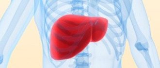
Interestingly, belly fat can cause a lot of problems, including fat that we don't actually see. This fat accumulates in the form of internal layers and surrounds the internal organs. Overweight people have a lot of fat between the internal organs. In this case, sometimes the liver becomes so “packed in fat” that it leads to a certain form of hepatitis; in special cases, it fails altogether, experts say. The good news is that eating right not only helps control the appearance of visible fat, but also prevents the inner layers of fatty tissue from appearing.
Fact or Fiction: Foods high in water-soluble fiber cause bloating and gas in the intestines, while foods high in insoluble fiber usually do not cause these problems.
This is true.
Fiber is dietary fiber found in most plants. It is the basis of cell walls in plant organisms. Fiber plays an important role in the functioning of the body, promoting good digestion. Many people don't know that there are different types of fiber. Water-soluble fiber is found in foods such as oatmeal, legumes, peas and citrus fruits - these foods are more likely to cause bloating and gas than foods with insoluble fiber - whole wheat bread, wheat, cabbage, beets and carrots.
Gas and bloating occur because the intestinal flora needs to digest soluble fiber. Since insoluble fiber is not digested at all, but simply passes through the gastrointestinal tract, it does not interact with the flora, so gases do not form.
Fact or Fiction: To get rid of reflux disease (sour belching), just lose a little weight
This is true.
The less acid enters the esophagus, the fewer problems. You may not believe it, but it’s enough to remove an extra kilogram from the stomach, and the result will be immediately felt.
During pregnancy, the baby grows and puts pressure on the internal organs, this can cause heartburn, but after childbirth, when the pressure goes away, the heartburn goes away.
The good news, experts say, is that if you lose weight first, you'll be free of heartburn within a few weeks.
Fact or Fiction: If you eat at night, you will gain weight faster than if you eat the same thing during the day
It is a myth.
Most experts believe that we gain weight because we consume more calories than we expend. And although it seems quite logical that the food we eat during an active day burns faster and is more effective than the food we eat before bed, weight gain is not at all dependent on the time of day. Whether you gain excess weight or not depends on how efficiently you spend your calories.
Recent animal studies have shown that avoiding eating in the evening may not help you lose weight. Eating at night can disrupt our body's circadian rhythms, altering the hormones that control appetite, causing us to gain weight.
Also, if you are tired or stressed, digestion is difficult before bed and you may experience bloating, gas, or heartburn. Our digestive canal has its own “brain” that helps food move through the digestive system correctly and in the right quantity. If we are tired, and this happens to almost everyone after a whole day of work, the “brain” of our intestines also gets tired, so it reduces the number of contractions, this, in turn, does not allow food to be properly digested.
Fact or fiction: Cookies with 200 kcal butter will control your appetite more than 200 kcal cookies without butter
This is true
. The reason is that fats are digested much more slowly than carbohydrates and stay in the stomach longer, which means we'll feel full longer if we eat a buttery cookie.
Moreover, simple carbohydrates (cookies, bread or pastries) are known to quickly increase blood sugar and insulin levels, which quickly decrease, leading to mood and appetite swings. You'll get hungry quickly.
Fact or Fiction: Legumes cause gas in everyone, and there's nothing you can do about it
It's a myth...sort of.
Legumes contain a lot of sugar, which requires a certain enzyme to be digested. Some people have a lot of this enzyme, others have a little. The less enzyme you have, the more gases will form in the intestines after eating legumes.
We have given only some of the questions that are often encountered by gastroenterologist patients.
We will be happy to answer your other questions in personal communication - Pirogova Irina Yurievna
Functions of the organ
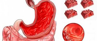
The main purpose of the stomach is to digest ingested food, and saliva, pancreatic juice and other secretions from the digestive tract contribute to this. After chewing, which occurs in the mouth, it goes through the esophagus to the stomach, where, under the influence of mechanical (peristaltic) muscle movement and the chemical action of hydrochloric acid, bile and other enzymes, it is converted into chyme. This mushy mass rises through the digestive tract to continue assimilation of essential nutrients and the resulting waste that cannot be used by the human body.
In the photo you can see a cross-section of the stomach
A secondary, but no less important function of the stomach is protective. Along with food, pathogenic bacteria also enter the body. Hydrochloric acid minimizes their number, that is, destroys them.
Pathology of the stomach and esophagus on ultrasound
Types of pathological changes in the stomach on ultrasound
Despite the difficulties of visualizing the stomach on ultrasound, due to the presence of a cavity in the organ, the accumulation of gases and the constantly changing shape due to peristalsis, the sonography method allows one to obtain fairly extensive and high-quality information about the condition of the stomach, on the basis of which various pathologies are identified.
Ultrasound signs of gastric pathology are the following:
- Changing the shape of an organ;
- Changes in the diameter of the stomach lumen;
- Changing wall thickness;
- The presence of irregularities on the inner or outer surface of the stomach wall;
- The presence of bulges on the wall of the stomach;
- Lack of peristalsis;
- Presence of antiperistalsis.
The main sign of stomach pathology is thickening of its walls and expansion/narrowing of the lumen of the organ. Thickening of the walls of the stomach with a characteristic uneven echogenicity, when the outer part of the wall is hypoechoic compared to the hyperechoic inner part, is called a symptom of damage to a hollow organ. Currently, in the world medical literature, the terms “target”, “lesion halo”, “pseudo-kidney”, “bull’s eye”, “pathological type of cockade” are also used as synonyms for the term “symptom of lesion of a hollow organ”. All of the above terms refer to the same pathological condition of the stomach, and therefore can be used as equivalent. However, in the countries of the former USSR, the most commonly used term is “symptom of an affected hollow organ,” which we will also use. Thus, thickening of the stomach wall, as a symptom of an affected hollow organ, can be diffuse and limited. With diffuse thickening, ultrasound shows an increase in wall thickness along its entire length. And with limited thickening, the wall of the stomach turns out to be thicker than normal only in a certain limited area. Unfortunately, the two main pathological signs of stomach diseases detected by ultrasound, namely the symptom of the affected hollow organ and the expansion/narrowing of the organ lumen, are nonspecific. This means that these pathological signs are inherent in many diseases, as a result of which it is impossible to accurately diagnose using ultrasound data alone. In order to correctly diagnose a person’s existing stomach disease and choose one of the many pathologies that are characterized by a symptom of an affected hollow organ or a change in the lumen of the organ, it is necessary, in addition to the ultrasound results, to also evaluate the clinical symptoms that worry the patient. Only a combination of pathological changes on ultrasound with clinical symptoms will help make the correct diagnosis. Below, for general orientation in the problem, we will provide a table in which we will indicate which diseases of the stomach are characterized by the presence of symptoms on ultrasound of the affected hollow organ and changes in the lumen of the organ.
| Diseases characterized by an ultrasound symptom of an affected hollow organ with diffuse wall thickening | Diseases characterized by an ultrasound symptom of an affected hollow organ with limited wall thickening | Diseases that are characterized by narrowing or widening of the lumen of the stomach cavity on ultrasound |
| Swelling of the stomach wall | Pyloric stenosis | Scar stricture |
| Chronic gastritis type B | Ulceration of carcinoma | Inflammatory stenosis |
| Ménétrier's disease | Stomach ulcer | Tumor-induced stenosis |
| Infiltrative carcinoma | Varicose veins | Impaired evacuation of gastric contents |
| Malignant lymphoma | Gastric carcinoma | |
| Malignant lymphoma of the stomach | ||
| Initial stage of malignant tumor | ||
| Benign tumors (leiomyoma, neuroma, stromal tumors) |
Swelling of the stomach wall can be observed in acute pancreatitis, nephrotic syndrome, congestive heart failure, as well as protein deficiency in the body. Pyloric stenosis can be congenital or acquired due to inflammation, ulcers or tumors.
Ultrasound picture for various diseases of the stomach
Currently, there are six main pathological processes of the stomach that can be accurately diagnosed using ultrasound - these are erosive and ulcerative lesions, polyps, pyloric stenosis, duodenogastric reflux, malignant tumors and malformations. Below we present the characteristic features of the ultrasound picture for the six different above-mentioned pathologies of the stomach, as well as damage to the organ.
Ultrasound picture of the developmental defect “pyloric stenosis”
With congenital pyloric stenosis, a baby develops persistent vomiting of gastric contents at 2–4 weeks of life. Ultrasound shows a pronounced symptom of the affected hollow organ, namely: sharp thickening of the walls in the antrum, narrowing of the walls in the central part and the general appearance of the stomach as an “hourglass”, as well as the presence of contents in the stomach more than 24 hours after ingestion. Ultrasound picture of the developmental defect “duplication of the stomach”
Using ultrasound, it is possible to establish a doubling of the stomach only by performing a water-siphon test so that both cavities are filled with water and the doctor can clearly see them. A diverticulum is an area of protrusion of the wall of the organ. Such diverticula can be single or multiple; on ultrasound they are visible only during a water-siphon test, when their lumen is filled with water and makes them visible. A diverticulum filled with water is an anechoic formation of any shape, bulging away from the wall of the stomach and connected to its cavity by a thin leg. Damage to the stomach usually occurs due to mechanical impacts on the area where the organ is located, for example, from blows to the stomach, a fall from a height onto the stomach etc. Such damage to the stomach can manifest itself as bruises, tears and ruptures of the walls of the organ. Less commonly, there are injuries caused by chemical substances, the picture of which on ultrasound is different from mechanical injuries. Gastric contusion is characterized by limited weakly echogenic thickening or bulging of the wall in the area of damage. The bruise is better visible when performing a water-siphon test than on an empty stomach. A rupture of the stomach wall is characterized by an uneven contour of the walls and their interruption in the area of \u200b\u200bthe rupture. When performing a water-siphon test, a rupture is easily diagnosed, since an ultrasound immediately shows the place where water leaks into the abdominal cavity. Chemical damage to the stomach wall is characterized by its thickening, doubling of the contour (an echo-negative stripe on the outside, and an echo-positive stripe on the inside). If, as a result of the damage, a cicatricial narrowing of the stomach cavity has developed, then an ultrasound will reveal fluid and food debris taken more than 14 hours ago. A gastric ulcer on an ultrasound looks like a hyperechoic thickening of the wall with its deformation and narrowing of the lumen. In addition, an excess amount of fluid on an empty stomach is recorded, a symptom of damage to a hollow organ. The following sizes of various parts of the stomach according to ultrasound are characteristic:
- Outer diameter 16 – 22 mm;
- Thickening of the wall of the gastric outlet from 6 to 10 mm;
- The distance between the walls in the thickening area is 7 – 12 mm;
- Image ratio 0.5 – 3.3 mm.
When performing a water-siphon test for a stomach ulcer, the following symptoms are characteristic:
- A clear, uneven contour of the wall in the area of the ulcer with hyperechoic inclusions;
- Uneven thickening of the internal hyperechoic layer of the stomach up to 2 – 4 mm;
- The wall thickness in the area of ulcer or erosion is always more than 5 mm;
- Indistinct layering of the wall in the area of the ulcer;
- Inactive peristalsis.
Ultrasound picture for gastritis
The walls of the stomach are evenly thickened, their echogenicity is uneven, with alternating hypoechoic and hyperechoic areas, more than 40 ml of liquid is detected in the cavity on an empty stomach, the diameter of the pylorus is more than 10 mm, signs of duodenogastric reflux. Stomach peristalsis is sluggish. Menetrier's disease is a giant hypertrophic gastritis, in which the gastric mucosa forms huge folds with polyp-like growths. Ultrasound shows a highly thickened wall with uneven echogenicity. Peristalsis is sluggish. A large amount of liquid and food taken more than 14 hours ago is found in the stomach. A characteristic symptom of damage to a hollow organ is with a thickening of the wall in the area where the polyp is localized to 20–28 mm, a gap between the walls in the area of the thickening of 3–12 mm. There is also an uneven structure of the stomach wall in the area of thickening, the image coefficient in the area of the polyp is 0.9 - 9. The amount of fluid in the stomach on an empty stomach is normal. When performing a water-siphon test on ultrasound, a polypoid bulging of the wall into the lumen of the stomach is clearly visible. The structure of the polyps is heterogeneous, there is no layering.
Ultrasound picture for malignant tumors of the stomach
There is always a symptom of damage to a hollow organ above the area where the tumor is localized. The wall is thickened unevenly (from 10 to 30 mm), the thickening area is hypoechoic, heterogeneous, with impaired layering. The outer contour of the stomach wall in the area where the tumor is located is uneven, but clear. The lumen of the stomach in the tumor area is 2 – 16 mm. The lower border of the greater curvature of the stomach is lower than normal. The amount of fluid in the stomach on an empty stomach is either within normal limits or slightly increased. When performing a water-siphon test, uneven filling of the cavity with water and its insufficient expansion are noted. Rigidity of the thickened wall and sluggish peristalsis are also characteristic. The internal contour of the stomach is hyperechoic, fragmented and thickened more than 3 mm. In the area of the tumor, the outer and inner contours of the stomach are uneven, but clear. Pyloric stenosis is a narrowing of the outlet of the stomach, due to which the evacuation of food masses into the duodenum is impaired. The causes of pyloric stenosis are usually tumors or a long course of a peptic ulcer, as a result of which scars are formed that tighten the walls of the stomach and reduce the lumen of its pyloric part. Pyloric stenosis is characterized by a shapeless hyperechoic formation with a sharp narrowing of the lumen of the affected area. At the initial stage, the walls of the stomach are thickened, but then they become thinner and the cavity expands. An ultrasound shows that the stomach is dilated and filled with contents of different ages. The amount of fluid on an empty stomach is more than normal, and there is sediment in it. The lower limit of the greater curvature of the stomach is significantly lower than normal. Peristalsis is either absent or weak. In the area of stenosis there is a symptom of damage to a hollow organ, and the wall is thickened to 7 - 22 mm, it has no layering, and the image coefficient is 0.6 - 8.5. When performing a water-siphon test for pyloric stenosis, the patient can only drink a small amount of liquid – no more than 300 ml, due to a feeling of fullness in the abdomen, nausea and sometimes vomiting. When liquid enters the stomach, its contents are mixed and sediment floats to the surface. The lower border of the greater curvature drops even lower than when examined on an empty stomach. Peristalsis is sluggish or absent. It is impossible to visualize the normal layering of the stomach wall in the area of stenosis. Duodenogastric reflux is a pathological condition in which the contents of the duodenum are refluxed into the stomach. Reflux is detected during a water-siphon test, and on an empty stomach it can be seen on an ultrasound only if there is an accumulation of fluid in the patient’s stomach in an amount greater than normal. On ultrasound, reflux is visible by the retrograde movement of hyperechoic particles from the duodenum up the stomach after filling it with water. The presence of fluid in the stomach on an empty stomach in an amount of more than 40 ml may indicate a peptic ulcer, gastritis, pyloric stenosis or a malignant tumor. In order to understand exactly what disease the fluid in the stomach indicates in a particular case, you need to take into account other signs on ultrasound, as well as the clinical symptoms that the person has.
Pathological changes in the esophagus and ultrasound images in various diseases
Pathological changes in the esophagus on ultrasound are changes in the diameter, wall thickness, length of the abdominal organ, as well as the angle of His. Let's consider what ultrasound signs are characteristic of various pathologies of the esophagus. GER is a pathological reflux of stomach contents into the esophagus. And GERD is damage to the lining of the esophagus due to reflux. GER and GERD are characterized by expansion of the lumen of the esophagus on ultrasound and thickening of its wall. Chalazia is weak peristalsis, as a result of which food stagnates in the esophagus, moving only under the influence of gravity. On ultrasound, chalazia of the esophagus is characterized by expansion of the lumen of the organ and thickening of its wall. Chalazia cardia is characterized by a gaping of the distal esophagus up to 6–14 mm, as well as visible flow of stomach contents into the esophagus. Cardiospasm is a pathological increase in the tone of the esophagus, as a result of which food does not move well to the stomach. Cardiospasm on ultrasound is characterized by expansion of the thoracic esophagus and at the same time a sharp narrowing of the esophageal-gastric junction to 3 - 5 mm. Stagnant contents are visible in the lumen of the esophagus, the wall of the organ is slightly thickened.
How does my stomach hurt?
The pain can be dull, sharp, paroxysmal, occur in the middle, above or below, on the right or left side of the body.
The intensity of such sensations does not always reflect the severity of the condition causing them, just as vice versa - mild but constant pain can be a sign of a chronic disease that needs urgent treatment.
If discomfort in the epigastrium persists for more than two weeks, you need to inform a gastroenterologist about this. This will avoid a more serious diagnosis. The reason to immediately consult a doctor is pain in the peritoneum, accompanied by constant bloating, regular vomiting, diarrhea and blood in the stool.
Diagnostic methods
It is possible to understand the cause of stomach pain and determine the correct treatment tactics based on instrumental and laboratory studies. At the initial examination, the localization and nature of pain is determined by palpation, as well as the degree of its irradiation.
Next, a set of studies may be prescribed:
- Ultrasound - determines the condition of the organ wall;
- MRI, CT are more informative methods that are necessary if tumors are suspected;
- clinical and biochemical blood tests;
- urine and stool tests;
- probing of the stomach and duodenum with further examination of the contents (acidity is determined, microscopy and bacterial culture are performed).
Diagnosis begins with simpler methods that allow you to identify the most common stomach diseases. If necessary, additional studies are prescribed. The Clinical Institute of the Brain has all the conditions for conducting a comprehensive diagnosis of stomach pain.
Accurate diagnosis
Identification of provoking factors that cause stomach pain always begins with a questioning of the patient and a physical examination - palpation of the abdomen, listening to the heart rhythm and lungs. After this, laboratory tests are carried out:
- Blood test (general and biochemical).
- Study of biomaterial (stool, urine and gastric juice).
To clarify the diagnosis, instrumental tests are also necessary - endoscopy, ultrasound, X-ray, computed tomography or magnetic resonance imaging of the abdominal organs with a contrast agent. In extremely rare cases, it may be necessary to conduct an examination using a laparoscope. When a flexible probe with a microcamera is inserted into the organ through a small incision for a detailed study and assessment of the general condition.
The location of the stomach explains the diversity of the nature of pain and such an extensive list of pathologies that cause them. Therefore, such a symptom should never be neglected. If you experience discomfort in the epigastrium, you must always seek medical help. Timely treatment in many cases guarantees recovery.