Causes
The main causes of hypercalcemia are excess parathyroid hormone in the body (hyperparathyroidism), oncology and long-term use of calcium supplements. Hypocalcemia almost always develops against the background of parathyroid hormone deficiency, the production of which is responsible for the upper and lower parathyroid glands. By interacting with the hormone calcitonin (thyroid), the exchange of phosphorus and calcium in the body is regulated. Excess calcium, as well as deficiency, develops with deviations in the functioning of organs and systems.
Where to get diagnosed and treated for hypocalcemia in Moscow
Make an appointment with an endocrinologist at the El.En clinic. The specialist will conduct an examination and prescribe the necessary diagnostic procedures. Tests can be carried out in the clinic; we have modern equipment for making a diagnosis. When making an appointment, you choose a convenient time.
The disease is treated through drug therapy; the patient is prescribed drugs that compensate for the lack of necessary substances - calcium, vitamin D, magnesium, etc.
At the first consultation you will receive answers to all questions regarding this pathology. Call us or leave a request on the website.
Symptoms
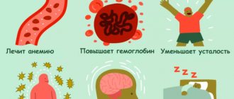
The first signs of hypercalcemia may not be noticeable, and only a random blood test will indicate problems. The appearance of obvious symptoms of increased calcium in the blood depends on the duration of this condition, the rate of development, and the severity of the underlying disease. These symptoms manifest themselves in the main systems of the body: nervous, muscular, digestive, urinary, cardiovascular, eye and skin diseases. The patient's memory deteriorates, lethargy, drowsiness, depression, weakness and muscle pain, restless leg syndrome at night, loss of appetite, constipation, nausea, gag reflex and belching appear. Possible weight loss due to pancreatitis, cholelithiasis, stomach ulcers with high acidity. Arthritis, arthrosis, and gout develop. Hypocalcemia is often combined with a lack of potassium, which leads to excessive excitability of neurons. As a result, muscle spasms occur (shoulders, hands, larynx, facial muscles). Skin sensitivity is also impaired, and a burning sensation or numbness occurs. Hemorrhagic syndrome develops, which is manifested by increased bleeding. Blood clotting decreases. Dystrophic changes in tissues, dental defects, brittle nails, dull hair, dry skin occur, heart rhythm is often disturbed, and cataracts develop.
Hypercalcemic crisis
A rare but dangerous complication is hypercalcemic crisis. In an acute condition, the patient’s entire nervous system is disrupted and blood clotting accelerates. Increased blood density implies increased risks for the patient’s life, as this can lead to the formation of blood clots or complete cardiac arrest.
Hypercalcemic crisis has symptoms:
- maintaining temperature above 40°C;
- internal bleeding;
- signs of fever;
- severe itching of the skin.
Disturbances of the central nervous system lead to the occurrence of psychosis, and subsequently shock. The patient's condition leads to the fact that he does not understand what is happening. If medical assistance is not provided in a timely manner, respiratory paralysis begins, followed by cardiac arrest and death.
Treatment
The initial stage of hypercalcemia, which is caused by excessive consumption of foods and medications containing calcium, can help solve the problem of a slight increase in the amount of calcium in the human body: changing the diet towards reducing the amount of foods containing large amounts of calcium. Dose adjustment, replacement or refusal of medications containing calcium. For healthy kidneys, sufficient consumption of water, preferably distilled (no more than 2 months). If hypercalcemia develops against the background of serious illnesses, then treatment is aimed at the underlying disease and cleansing the body of excess calcium. When the kidneys are functioning normally, calcium is usually washed out with the help of diuretics and intravenous saline. In severe conditions, hemodialysis is performed (cleansing the blood of waste products). If the process of increased removal of calcium from the bones cannot be stopped, then hormonal drugs are used. The main task of hypocalcemia is to compensate for calcium deficiency in the body. In addition, treatment is aimed at eliminating the cause of the disease. So, for hypoparathyroidism (lack of parathyroid hormone), hormonal therapy is prescribed. Chronic hypocalcemia is treated by regularly taking calcium tablets and vitamin D. In addition, measures are taken to normalize the levels of magnesium, potassium and protein in the blood.
Hypocalcemia
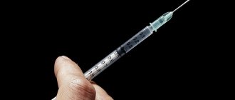
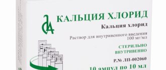
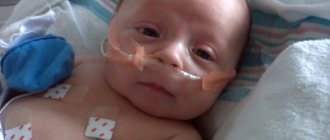
etiology, pathogenesis Hypocalcemia and hypocalcemic tetany (HT) are a striking metabolic-endocrine syndrome, the leading clinical manifestations of which are paresthesia and generalized, and sometimes local, convulsions associated with a decrease in calcium concentration in the blood. HT is observed in 70–90% of patients in intensive care units due to pancreatitis, sepsis, major operations and injuries, as well as other acute conditions [1]. At the same time, HT is often recorded as the first and even the only manifestation of various chronic or latent pathologies [2, 3]. Numerous etiological and pathogenetic variants of HT are given in the classification. Classification of hypocalcemia: • Dysregulation of calcium-phosphorus-magnesium metabolism. I. Parathyroid hormone deficiency, i.e. hypoparathyroidism (HyPT): 1) removal or damage of the parathyroid glands (PTG) during surgery; 2) radioiodine therapy or radiation therapy for diseases of the neck organs; 3) hemochromatosis; 4) tumor metastases in the parathyroid gland; 5) congenital underdevelopment of the parathyroid gland (idiopathic HyPT); 6) autoimmune destruction of the parathyroid gland. II. Violation of the action of parathyroid hormone: 1) pseudohypoparathyroidism (Phypoparathyroidism) or Martin-Albright syndrome: 1. PhiPT type 1; 2. PGIPT type 2; 2) pseudohypohyperparathyroidism (Costello-Dent syndrome). III. Impaired secretion or action of other hormones that regulate calcium-phosphorus-magnesium metabolism: 1) excessive secretion of calcitonin: 1. medullary thyroid cancer; 2. other apudoms; 2) vitamin D deficiency or impaired action of D-hormone: 1. rickets or osteomalacia in adults; 2. malabsorption syndrome; 3. insensitivity to vitamin D. • Functional hypocalcemia: 1) alkalosis (respiratory, metabolic, iatrogenic); 2) hyperproteinemia (hyperalbuminemia); 3) “hungry bones” syndrome; 4) neonatal hypocalcemia of infants born to mothers with hyperparathyroidism; 5) increased calcium uptake by osteoblastic tumors; 6) chronic renal failure (CRF); 7) endocrinopathies (diabetes mellitus, hypogonadism, hypopituitarism); acute destructive pancreatitis; 9) rhabdomyolysis. • Toxigenic and iatrogenic hypocalcemia: 1) excessive intake of phosphorus into the body; 2) hypomagnesemia; 3) introduction of EDTA; 4) treatment with mithramycin, neomycin, cisplatin; 5) use of phenobarbital, phenytoin, glucagon, laxatives, antacids; 6) massive infusion of citrated blood or extracorporeal hemoperfusion. By definition, the pathobiochemical factor of HT is a low concentration of circulating and cytosolic calcium, which is accompanied by increased excitability of neuromuscular and interneural synapses and, as a consequence, paresthesia, muscle cramps and other neuroregulation disorders. The most common cause of HT is hypoparathyroidism (HyPT), which complicates approximately 8% of surgical operations on the thyroid gland and almost all interventions on the parathyroid gland [4, 5]. Thyroid surgery, even by experienced surgeons, is fraught with the development of hyperthyroidism, since in approximately 2% of people the PTG is located inside the thyroid gland [6]. Irreversible HyPT sometimes occurs after radioiodine therapy for thyrotoxicosis, thyroid cancer, or radiation therapy for other malignant tumors of the neck [2, 3]. Destruction of the parathyroid gland by tumor metastases is considered a casuistic cause of hyperthyroidism and tetany. Meanwhile, such cases are apparently not uncommon, because some authors found metastases in the parathyroid gland in 5–12% of patients with cancer pathology, especially in breast cancer [2, 3, 5]. Autoimmune HyPT as part of autoimmune polyglandular syndrome (APS), like the syndrome itself, is an undoubted rarity, and congenital aplasia or hemochromatosis of the parathyroid gland is simply a casuistry. There is only one report of HyPT due to hemochromatosis of the parathyroid gland after multiple infusions of iron supplements for hypochromic anemia [1, 5]. Idiopathic pseudohypoparathyroidism (PhyPTH) is the insensitivity of target organs to parathyroid hormone (PTH). There are two forms of the syndrome, differing in that with PhiPT-1, the injection of parathyroid hormone does not affect the excretion of cAMP and phosphaturia, and with PhiPT-2, after the administration of PTH, the excretion of cAMP increases, but phosphaturia does not (Table 1). Pseudohypohyperparathyroidism is a variant of PhiPT, in which the kidneys are resistant to PTH, but skeletal PTH receptors are preserved. As a result, a bone form of primary hyperparathyroidism (osteitis fibrosis cystica) is formed, but calcium in the blood is not increased, but decreased [4, 5]. Pseudopseudohypoparathyroidism is another genetically determined syndrome, but here the defect is not in the PTH receptors, but in the PTH molecule [4, 5]. The syndrome is recognized by a test with PTH, during which both cAMP excretion and phosphaturia increase (Table 1). In terms of its effect on calcium-phosphorus-magnesium metabolism, calcitonin (CT) is an antagonist of PHT and D-hormone. In this regard, it is logical to expect that medullary thyroid carcinoma and other APUD cell tumors that secrete a lot of CT should be accompanied by attacks of tetany. However, in practice, hypocalcemia is extremely rare in such patients, which is explained by the powerful effect of PTH and D-hormone, which block the effects of calcitonin. The pathogenesis of tetany with vitamin D deficiency or a defect in D-hormone receptors is close to that with HyPT and PhiPT. This makes their differential diagnosis difficult, which, however, is possible using a PTH test. After eliminating hypocalcemia by administering calcium, patients with D-hormone deficiency give a normal phosphaturic response to the administration of PTH, while in HyPT and PhiPT, parathyrine does not increase phosphaturia. Hyperalbuminemia and alkalosis reduce the level of ionized calcium, which, in combination with other causes, can cause HT. Hungry bone syndrome is the name given to hypocalcemia that often occurs after surgical treatment of severe thyrotoxicosis or hyperparathyroidism. Temporary hypocalcemia in these cases is due to the fact that bone tissue intensively absorbs calcium, which has been “washed out” of the bones by excess thyroid hormones or PTH. Neonatal hypocalcemia of infants born to patients with hyperparathyroidism is explained by transient hypersecretion of CT and suppression of parathyroid function in infants against the background of excess parathyrine and calcium in the blood of the mother. The mechanism of development of hypocalcemia in patients with massive osteoblastocytomas that absorb calcium is obvious. Hypocalcemia in chronic renal failure is associated with increased loss of calcium in the urine due to a decrease in renal Ca2+ reabsorption and insufficient synthesis of D-hormone in the kidneys. Hypocalcemia against the background of some endocrinopathies is explained by a weakening of the permissive or increased opposing effect of systemic hormones in relation to PTH, CT and D-hormone. Finally, hypocalcemia in pancreatitis and rhabdomyolysis is due to chelation, i.e. calcium deposition in areas of massive destruction of adipose and muscle tissue, as well as hyperphosphatemia and hyperkalemia, which increase calcium excretion. In addition, these patients have impaired renal, gastrointestinal and endocrine system function. The phosphate ion (PO3-) is reciprocally bound to the calcium ion, so hyperphosphatemia is necessarily accompanied by hypocalcemia. This, in particular, explains late postnatal tetany and iatrogenic rickets if the child is not breastfed, but cow's milk, which contains a lot of phosphorus. The nature of hypocalcemia against the background of hypomagnesemia is not entirely clear, just as the nature of hypomagnesemia itself is not always clear. It is believed that hypomagnesemia inhibits PTH secretion and receptor binding [7]. The most common causes of hypomagnesemia are alcoholism, cytostatics, diabetes mellitus, parenteral nutrition, and a congenital defect in the intestinal absorption of magnesium. Iatrogenic hypocalcemia from a number of drugs is due to their chelating effect or blockade of vitamin D, or inhibition of intestinal calcium absorption. HT syndrome often complicates massive citrated blood transfusion or cardiopulmonary bypass procedures [3]. The sodium hydrogen citrate contained in preserved blood converts Ca2+ into poorly dissociated calcium hydrogen citrate. Clinical picture and diagnosis of hypocalcemia The most striking manifestation of hypocalcemia is prolonged, usually generalized, tonic convulsions, the so-called tetany. Tetany is painful for the patient, but the danger to life is low. In a few reports, death from tetany in the setting of postoperative hypoparathyroidism has been associated with asphyxia from laryngeal stridor and diaphragmatic contraction or cardiac tetany [1–3]. However, chronic hypocalcemia creates serious problems for the patient's health. It is associated with the development of cataracts, metastatic calcifications in organs and the brain, severe mental disorders with depression and suicide, infertility or miscarriage with fetal death, and finally, against the background of hypocalcemia, a chronic infection is activated. The classic triad of incipient HT is a combination of paresthesia, difficulty breathing and convulsions. Paresthesia begins from the lips and spreads to the hands and feet. Tonic convulsions can be local - a sardonic mask on the face or “carpopedal syndrome”. Almost always, seizures are accompanied by clear neurological symptoms - dysarthria, dysphagia, paresis of cranial nerves, stigmata of cerebrovascular, extrapyramidal and brainstem disorders, spastic paresis of the limbs. Disorders of the somatic nervous system are accompanied by dysfunction of the autonomic nervous system: profuse sweating, bronchospasm, renal or hepatic colic, vomiting, diarrhea. Intracranial hypertension and swelling of the optic nerves are often recorded. Convulsive syndrome in patients with HT is often regarded as epilepsy, although in epilepsy the seizures are usually clonic, with loss of consciousness. However, HT can also occur in the form of clonic convulsions with syncope, i.e. with short-term loss of consciousness. Sometimes HT resembles mental disorders: paranoid or hallucinatory psychosis, depressive-catatonic attack. Manifestations of hypocalcemia such as laryngospasm, tetany of the stomach and heart were mentioned above. In this case, asphyxia or “acute abdomen” syndrome, or an attack of angina pectoris and even small focal myocardial infarction are attributed, because The ECG shows QT prolongation, ST depression, sharpening or inversion of T. It is clear that the listed phantoms can cause therapeutic and diagnostic errors, sometimes dramatic, for example, hospitalization in a psychiatric clinic or surgery. Atypical and latent HT are recognized using tests for spasmophilia: • Khvostek's sign: twitching of the lips or contraction of the orbicularis oris muscle when tapping with a fingertip in front of the tragus of the auricle or between the zygomatic arch and the corner of the mouth. • Weiss's sign: contraction of the facial muscles when tapping on the outer edge of the eye orbit (outer corner of the eye). • Trousseau's symptom: the appearance of the so-called “obstetrician's hand” after compression of the arm with a sphygmomanometer cuff. • Schlesinger's symptom: in a patient lying on his back, with passive flexion in the hip joint, the thigh muscles contract convulsively and supination of the foot is noted. It must be remembered that symptoms of spasmophilia are present in 10–25% of healthy people and in all patients with vegetative dystonia and psychasthenia. At the same time, 30% of patients with hypocalcemia do not have signs of spasmophilia [1, 3]. In the diagnosis of hypocalcemia, anamnesis is important: previously observed convulsive attacks, thyroid surgery, radioiodine therapy or radiation therapy to the neck, chronic renal failure, enterocolitis, frequent fractures. There are specific signs for certain types of chronic hypocalcemia. For example, postoperative hypoparathyroidism, in addition to a scar on the neck, is characterized by eczema and other dermatoses, dry skin, hard, dry and brittle hair, uneven, brittle nails. Cataracts are often detected, which are considered a specific complication of chronic hypocalcemia. Ro-graphy reveals calcification of the basal ganglion, calcifications in the subcutaneous tissue, muscles, internal organs, subperiosteal ossification of tubular bones, diffuse fibrous osteitis [8]. Pseudohypoparathyroidism in 75% of cases has clear morphotypic stigmas: short stature, round face, short neck, brachydactyly, rachitic skeletal deformities, mental retardation [2, 5]. A screening test for hypocalcemia is the determination of calcium in the blood. The lower limit of total calcium is 2.2 mmol/l, ionized calcium is 1 mmol/l. The diagnosis is clarified using additional tests (Table 1). Treatment of hypocalcemic tetany Standard emergency therapy for HT is an intravenous bolus injection of 10–20 ml of a 10% calcium chloride solution. You need to beware of extravasal ingestion of calcium chloride, because it causes massive tissue necrosis. The danger of extravasation is very high, since the injection has to be done against the background of seizures. Instead of calcium chloride, you can use a 10% solution of calcium gluconate or lactate, however, to relieve HT with these solutions, you need to administer at least 20 ml, and preferably 40 ml, since they contain half as much Ca2+ ion per unit volume. It should be remembered that massive extravasation of these drugs also causes tissue necrosis. The most reliable way to prevent such a complication is to administer calcium solutions not as a stream, but by infusion through a cannula in a vein of 50 ml of a 10% CaCL2 solution diluted in 100–200 ml of a 5% glucose solution. If the diagnosis of HT is beyond doubt, and the administration of calcium-containing solutions does not have an effect, you should think about alkalosis or hypomagnesemia. These conditions can be confirmed and eliminated ex curantibus - by intravenously administering 10 ml of a 5% solution of ascorbic acid and 10 ml of a 25% solution of magnesium sulfate. But it is better to clarify the diagnosis by determining the concentration of magnesium and blood pH. The introduction of 10–20 ml of 10% calcium chloride stops tetany for only 2–3 hours. Therefore, it is advisable to immediately administer 40 ml of 10% CaCL2, or better, as mentioned above, to carry out a long intravenous infusion of 50–100 ml of 10% calcium chloride, diluted in 200–400 ml 5% glucose. The algorithm for diagnosing and treating HT is shown in Figures 1 and 2. After stopping an attack of HT, calcium and vitamin D preparations are prescribed orally. Calcium preparations irritate the mucous membrane of the stomach and intestines, so their dose should be as low as possible, and it is advisable to abandon them as quickly as possible. The patient is recommended a diet enriched with calcium, including dairy products, hard cheeses, soy, sesame, almonds, dried apricots, celery, canned fish “with bones”: sardines, sprats, pink salmon. Normally, the daily requirement for Ca2+ is at least 1000 mg. Naturally, in patients with most forms of hypocalcemia, the need for calcium is 2–3 times higher. It is clear that it is almost impossible to provide 2000–3000 mg of Ca2+ from food products. Therefore, the “calcium diet” still has to be supplemented with calcium supplements, preferably twice a day. Of the numerous vitamin D preparations, its active metabolites are preferred: calcitriol and alfacalcidol, prescribed in a dose of 0.25–1.0 mcg 1 or 2 times a day. A calcium-rich milk-vegetable diet contains a lot of phosphorus, which enhances calcium excretion in the urine and its absorption by the skeleton. To bind phosphorus, take 20–40 ml of aluminum hydroxide during meals. If hypocalcemia is not effectively eliminated, magnesium deficiency should be assumed and appropriate medications should be prescribed. These are magnesium sulfate, potassium and magnesium aspartate, magnesium orotate, etc. The adequacy of treatment is monitored clinically (absence of symptoms of spasmophilia) and by monitoring the levels of calcium, phosphorus and magnesium in the blood. In conclusion, it should be noted that it is necessary to focus on the prevention of HT, which consists in the timely diagnosis of hypocalcemia, which is achieved by mandatory determination of calcium in the blood, better ionized, in all outpatient and hospital patients.
Literature 1. Marino P. Intensive care; lane from English M.: GEOTAR-Media, 1998. P. 461–463. 2. Lukyanchikov V.S., Korolevskaya L.I. Hypoparathyroidism and hypocalcemic syndrome. // Clinical medical 2003. No. 1. P. 66–70. 3. Carlstedt F., Lind L. Hypocalcemic Syndrome // Critical. Care Clinics. 2001. Vol. 17. No. 1. P. 139–153. 4. Potts J. Hypocalcemia // Internal diseases (translated from English) / Ed. E. Braunwald et al. M: Medicine, 1997. T. 9. pp. 397–411. 5. Wilson J., Foster D. Hypoparathyroidism. In: Williams Textbook of Endocrinology (8th ed.). . Philad.: WB Sounders, 1992. P. 1456–1476. 6. Wheeler M., Williams E., Path F. et al. The Hyperfunctioning intrathyroidal parathyroid gland // World. J. Surg. 1987. No. l. R. 10–114. 7. Dacey M. Hypomagnesemic disorders // Critical. Care Clinics. 2001. Vol. 17. No. 1. P. 155–179. 8. General Guide to Radiology (translated from English) / Ed. N. Pettersson. M.: RA “Spas”, 1996. T. 2. P. 690–733.
Diagnostics
Calcium metabolism disorders are determined by laboratory tests. To determine the cause of the pathology, more extensive research is required, so specialists prescribe hardware studies. Additionally, differential diagnosis can be carried out.
Examination methods:
- urine test (general) - with elevated Ca levels, a mineral will be present in the biomaterial and the alkaline reaction will be pronounced;
- blood test (biochemical) - a significant increase/decrease in ionized and total calcium is detected in the plasma;
- ultrasound examination - the entire endocrine system is examined for the presence of tumors, tissue enlargement and other abnormalities;
- radiography - used to examine bones to determine the possible development of osteoporosis, fractures, cystic formations and other pathological processes;
- densitometry - used to accurately determine the density of bone tissue;
- radiography (with contrast agent) - helps to identify ulcerative lesions of the gastrointestinal tract and parathyroid adenoma in the retrosternal space;
- computed tomography - examines the urinary system and kidneys for the presence of stones;
- magnetic resonance imaging - allows you to obtain the most accurate data about tumor formations of the thyroid glands;
- spintigraphy is a study using radioactive isotopes, with the help of which it is possible to visualize the area under study.
Diagnostics allows you to obtain the maximum amount of information to identify provoking factors, characteristics of the disease and possible complications. Treatment can begin only after receiving the examination results.