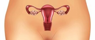
With this disease, areas of altered squamous epithelial cells (covering layer) form on the mucous membrane of the lower part of the uterus and vagina. Typically, dysplasia affects the vaginal area and the cervix at the same time, which complicates the situation. Epithelial dysplasia is classified as a precancerous pathology.
Appointment with a gynecologist - 1000 rubles. Comprehensive pelvic ultrasound - 1000 rubles. Extended colposcopy - 1300 rub. Appointment based on ultrasound or test results - 500 rubles (optional)
CLICK TO MAKE AN APPOINTMENT or ultrasound tests
What is dysplasia
The content of the article
On the Internet, this disease is often described as abnormal cell growth. In fact, the pathology is much more serious than this primitive description. WHO characterizes epithelial dysplasia as a complex of disorders, including:
- cellular atypia - modification or replacement of cells with unusual ones for a given tissue;
- failure in cell differentiation - genetic code responsible for cell functions, their size, shape and metabolism;
- failure in the architectonics of the fabric - structure, structure, etc.
Dysplasia is not only cellular atypia, it is deviations in the entire tissue complex.
Due to the asymptomatic nature of the disease in the first stages, treatment of vaginal dysplasia often begins late, when the pathological process covers most of the area of the mucous membrane of the cervix and vagina. Early vaginal dysplasia is usually diagnosed at a routine appointment with a gynecologist or during an examination related to another pathology.
Types of dysplasia
Dysplasias are very diverse and heterogeneous, congenital and acquired dysplasias are essentially very different processes, some involve several systems, others only areas of the mucous membrane.
Congenital dysplasias are manifested by developmental anomalies - deviations in the structure that do not interfere with normal functioning, and developmental defects - these anomalies already disrupt the functioning of the anatomical region.
Mucosal dysplasia does not form defects or developmental anomalies, but can lead to malignant processes. Absolutely different manifestations of dysplasia do not allow creating a unified classification; each researcher of the problem offers his own systematization of heterogeneous pathology, not free from shortcomings.
Congenital dysplasias are formed in the prenatal period; clinical manifestations affect the integumentary tissues and the musculoskeletal system; their division is very conditional.
Dysplasia of the ectoderm is distinguished - the superficial tissue of the embryo from which the outer integument is formed: skin with nails and hair and the oral mucosa with teeth. Pathology can manifest itself at any age, in any set of symptoms and with any severity. This can only be dry skin with small spots of atrophy and an almost complete absence of teeth, or baldness with no nails and moon-shaped defects in the tooth enamel with a full set of teeth. It is also possible that there is simply a decrease in the number of sweat glands in the skin, which leads to rapid overheating already in infancy and the danger of sudden death.
The vast majority of those suffering from genetic connective tissue dysplasia may experience disorders of all body systems from minor to life-threatening and with any set of symptoms: neurological disorders, heart valve defects, skeletal changes, vascular aneurysms, softening of the tracheal rings, prolapse of the kidneys, and also leading to sudden death due to combined damage to the cardiovascular and respiratory systems. The most active surge in manifestations is observed in adolescence, when, for example, scoliosis and flat feet progress sharply, cardiac arrhythmia and myopia appear. Several syndromes with characteristic external manifestations have been formed, again with variable severity - from scanty individual manifestations to extremely pronounced ones with early death from complications, such as Marfan syndrome and osteogenesis imperfecta.
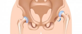
Joint dysplasia is manifested by congenital malformations of large - hip and knee, as well as small joints of the hand and foot, and in essence can be considered the same connective tissue dysplasia, but in a separate articular system. As a rule, the pathology is detected in infants, but in a minimal form it can go unnoticed, which is discovered during an examination for an accidental leg injury.
Fibrous dysplasia is indicated by single and multiple cysts in the bones. Some people live healthy lives into old age and learn about genetic pathology in the form of a small brush by chance during an X-ray for another reason. Others suffer from chronic pain and limb deformity, often with shortening, due to multiple cystic cavities in the bone tissue.
Dysplasia of the mucous membranes of internal organs is “earned” during life. In most cases, the process does not manifest symptoms or has uncharacteristic and unstable minimal signs, which are found by microscopy of a piece of tissue. Dysplasia can progress from mild, through moderate and severe, up to stage zero cancer - and this is its main danger. Severe dysplasia is difficult, and sometimes impossible, to reliably distinguish from non-invasive carcinoma in situ.
Gastric dysplasia can be clinically manifested by gastric pathology; the patient is often infected with Helicobacter. The pathology lies in the simplification of the cellular structure - decreased differentiation, changes in cell nuclei and other intracellular structures. Mild dysplasia not only turns into moderate, but also regresses; moderate dysplasia proceeds similarly, while severe dysplasia is considered a precancerous process and is subject to serious treatment and observation. Gastric dysplasia in frequency of occurrence is significantly inferior to metaplasia - the formation in the mucosa of areas of cells very similar to the cells of the large or small intestine. During microscopy, areas of intestinal metaplasia are always found around any dysplasia, and this is also a precancerous process.
Primary urothelial dysplasia is an uncommon pathology, manifested by symptoms of an irritable bladder with frequent and painful urge to urinate. This variant of dysplasia is not usually divided into degrees; the probability of developing cancer on a dysplastic background is slightly more than 15% and the transition to stage zero cancer will take from 4 to 8 years.
Cervical or dysplasia of the cervical mucosa is a very common pathology, since its leading cause is infection with the human papillomavirus. The virus itself cannot be killed - it is resistant to any drugs, but it is fatal and in the vast majority of women it will heal on its own in the next two to three years.
Causes of development of vaginal dysplasia
The main causes of cell degeneration are genetic predisposition and infection with oncogenic human papillomaviruses (HPV), HSV-2, HPV and HIV.
The development of dysplasia is accelerated by:
- Hormonal problems associated with menopause - lack of estrogen (hypoestrogenism), taking contraceptives without a prescription, etc.;
- Diseases that change the vaginal flora and weaken the immune system: vaginitis, colpitis;
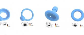
- Conditions that deplete tissue, for example, rubbing the vagina with a gynecological ring, using vibrators, low-quality condoms and tampons.
At risk are women with bad habits - those who smoke, consume fast food and alcohol, and live in areas with a poor environmental profile.
Mechanism of disease development
The connection between the presence of human papillomavirus (HPV) and cervical dysplasia has been established by a number of medical studies dating back to the pre-war years. The vast majority of people who are sexually active experience HPV infection. Most often, the body copes with it without medical help. Even precancerous stages in women with diagnosed dysplasia can be cured by the immune system. But the body’s capabilities do not guarantee such an outcome, therefore, given the huge risk of threat to health and life, it is necessary to undergo adequate treatment for the current stage.
The papilloma virus is transmitted sexually. Penetration is not necessary to acquire an infection - external contact of the genitals is sufficient. Most types of HPV do not cause adverse health effects and go away on their own. If there is no re-infection with the same type of virus, then after two years the infection is no longer recorded and does not manifest itself in any way in the body. But in some cases, HPV infection becomes chronic, causing cervical dysplasia with the risk of the formation of cancer cells. From the moment of infection to the cancer stage, with normal immunity, it can take 15-20 years. In HIV-positive patients who are not undergoing therapy, this period is reduced by 2-3 times.
Causes of cervical dysplasia
The most common cause of dysplastic changes in the cervix is infection with papillomavirus. HPV types 16 and 18 are found in 98% of cases of dysplasia. The disease is provoked by:
- inflammatory processes in the vagina;
- trauma to the cervix during childbirth and abortion;
- hormonal dysfunction;
- reduced immunity;
- alcohol and smoking;
- too early and promiscuous sex life;
- neglect of genital hygiene.
With dysplasia, the structure of the epithelium is disrupted, and the mucous membrane does not perform protective functions. With pronounced dysplastic changes, the woman is referred for consultation to an oncologist.
Symptoms - when to seek help
There are no clinical manifestations of first-degree cervical dysplasia; it can only be detected during examination. However, in most cases, the disease occurs against the background of other ailments that have certain symptoms. Associated inflammatory processes can manifest as burning or itching, discharge of varying consistency, and contact microbleeding after sexual intercourse. Frequent accompaniments of dysplasia may be gonorrhea or chlamydia, which have specific symptoms.
Statistics show that in most cases, dysplasia is accompanied by the presence of uterine erosion, so an experienced specialist will definitely refer you for a special PAP analysis in such cases. If cervical erosion is detected during pregnancy, the diagnosis of dysplasia can be carried out using conventional methods. The first and second stages are not an obstacle to normal childbirth; in the third stage, the doctor may recommend a cesarean section.
Stages of development of dysplasia
The dysplastic process is a continuation of hyperplasia - an increase in the number of cells caused by chronic inflammation and degeneration. Often hyperplasia and dysplasia are accompanied by tissue atrophy (death), since these processes have common genetic mechanisms.
To be precise, the term “dysplasia” is not used in medicine to characterize transient precancerous processes. With pathology in the vaginal sector of the cervix, the condition is designated CIN (cervical intraepithelial neoplasia), precancerous changes in the vagina are designated - VaIN, vulva - VIN.
There are three degrees of dysplasia:
- Mild, mild (D I)
– up to 1/3 of the thickness of the epithelial layer is affected; - Moderate, medium (D II)
- altered cells grow on 2/3 of the epithelial tissue; - Severe, pronounced (D III)
- the entire layer is changed. This degree of dysplasia is the initial stage of cervical and vaginal cancer.
The determining criterion for the degree of dysplasia is the severity of cellular atypia. The more severe the degree, the greater the size, hyperchromicity and polymorphism of the cell nuclei. Epithelial dysplasia can regress (reverse process), be stable or progress. How quickly the process of malignancy progresses depends on the severity and duration of the disease. The more significant the dysplasia, the lower the likelihood of regression.
Severe dysplasia is regarded by gynecologists as an obligate precancer that guarantees the development of cancer. Therefore, patients with obligate precancer are registered with an oncologist.
Kinds
Cervical dysplasia is classified according to severity depending on the volume of epithelium involved in the dysplastic process. There are three degrees of the disease. With grade 1 (weak, mild) neoplasia, minor tissue changes occur with a moderate proliferation of atypical cells (proliferation) of the basal layer. This stage is characterized by a morphological sign - koilocytosis (koilocytes - cells affected by HPV) and dyskeratosis (keratinization of the epithelium, characterized by the presence of scales).
With weak neoplasia, the pathological process extends to no more than one third of the depth of the epithelial layer from the basement membrane. The small area of the lesion makes it somewhat difficult to take a biopsy sample for morphological examination.
With the other two degrees, atypical changes are more pronounced. CIN 2 (second stage or moderate, moderate) is characterized by damage to 50% of the thickness of the epithelial tissue, while CIN 3 (third stage or severe, non-invasive cancer) is characterized by damage to more than two-thirds of the epithelium. The prognosis depends on the stage of the process; the most favorable prognosis is grade 1 of pathology.
Degrees of the disease
Symptoms of vaginal dysplasia
At the beginning of the disease, patients do not feel anything. Foci of dysplasia in the vagina are discovered accidentally during a gynecological examination. The only symptom indicating infection with the HPV virus (it causes pathology in 90% of cases), which the patient can see with the naked eye, is small warts (genital warts) in the genital area.
As the pathology develops, the following appear:
- redness and dryness of the vaginal mucosa;
- bleeding after intercourse or douching;
- discharge with an unpleasant odor;
- itching, burning and swelling of the vaginal area.
With severe dysplasia, pain in the sacrum and swelling of the legs occur. While walking, there is discomfort in the vaginal area.
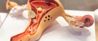
A gynecologist, conducting an examination using a colposcope (optical device), sees changes in the vaginal mucosa in the form of protruding reddish or light spots with uneven outlines. Dysplasia areas are large and affect the cervix. To clarify the diagnosis, the mucous membrane is lubricated with solutions of vinegar or Lugol (extended colposcopy). With advanced dysplasia, mosaic mucosa and papillary growths are visible.
The final diagnosis is made after a biopsy of suspicious areas, during which the gynecologist takes part of the cells for analysis.
Diagnostics
The most accessible and accurate method for diagnosing dysplasia and early stages of cervical cancer is a cytological study of a cell preparation (scraping of epithelial cells of the cervix). This method is also known as a PAP smear, named after the Greek physician Papanicolaou. The procedure for taking a smear is painless. The gynecologist uses a sterile instrument to scrape cells from the outer mucosa of the cervix. The taken material is sent to the laboratory, where it is examined under a microscope. During a microscopic examination, specialists determine the condition of the cells based on morphological characteristics. Healthy cells form a uniform epithelial structure, and their nuclei are small. Enlarged nuclei and scattered structure indicate the presence of the disease. The degree of dysplasia is determined by the morphology of the cells and the depth of epithelial damage.
The initial stage of dysplasia is invisible when examining the outer part of the cervix. Erosive manifestations are observed when damaged epithelial cells form lesions. They may appear as redness with a whitish coating. But the final diagnosis is made only after a biopsy.
A PAP smear allows you to detect the presence of atypical cells in the scraping. If such cells are identified, then a biopsy procedure is prescribed to clarify the diagnosis and determine the degree of dysplasia. The colposcope is inserted into the vagina. The optical system helps to view areas at high magnification. A tissue sample is taken from the most changed areas, usually from three points.
Of the two hundred HPV subtypes, only a few are classified as carcinogenic. To determine the strain, a vaginal smear is prescribed. Laboratory testing using the PCR (polymerase chain reaction) method allows you to detect and identify the DNA of human papilloma viruses. This is the most informative method for determining the causative agent of dysplasia.
Symptoms of cervical dysplasia
In this condition, cells appear in the cervical epithelium that differ in structure from normal ones. The process affects the basement membrane and the upper layer of tissue. The disease often does not produce symptoms, therefore, in order to recognize the disease in time, it is necessary to regularly do colposcopy and undergo tests. With severe dysplastic pathology, women complain of itching, pain and burning in the vagina. The discharge becomes yellowish or bloody. Contact bleeding is possible.
To confirm the diagnosis, colposcopy is performed with taking a smear from the cervix and cervical canal for histological and cytological examination. With dysplasia, a changed area is visible on the cervix, and dysplastic cells are found in the cervical mucus.
Postoperative period
Recommendations in the postoperative period depend on the type and volume of intervention that was performed. The woman should be at rest, which will promote faster tissue healing.
Doctors recommend that within 2-3 months after surgery you avoid:
- Moving heavy objects.
- Physical education classes.
- Visits to baths, saunas.
- Sexual contacts.
To monitor the healing process, it is important for a woman to regularly visit a gynecologist after each menstrual cycle. If bleeding does not stop within 6 weeks after the intervention, an unpleasant odor, itching and burning are observed, this indicates the presence of complications. In this case, the recovery period may be delayed, and the uterine cavity will become scarred.
How to cure vaginal dysplasia
Because of the term “precancerous condition,” patients, faced with vaginal or cervical dysplasia, become depressed. But in reality everything is not so scary!
Treatment depends on the degree of dysplasia:
- Mild dysplasia does not require treatment until the condition worsens. If the doctor notices changes, the modified cells are destroyed with a laser, chemicals or modern radio wave method. Genital warts are removed using the same method, as they can develop into cancer.
- In case of deep dysplasia or the onset of cancerous degeneration, vaginectomy is indicated - removal of dysplasia areas. If the process has gone too far, then to save the organ, skin is transplanted from the buttocks or thighs.
Pregnancy with grade 3 dysplasia
Among the most common complications during pregnancy against the background of grade 2 and 3 cervical dysplasia are the threats of miscarriage and premature birth. After surgical treatment of dysplasia, pregnancy can be planned no earlier than six months later. All this time, the woman should be observed by a gynecological oncologist.
Patients who have undergone surgery to remove areas of the cervix affected by dysplasia are at high risk of complications during pregnancy. Reproductive function is possible, but women with cervical surgery with an established viral origin of the disease should be under systematic medical supervision. Attention is paid primarily to the study of epithelial covers for the presence of new viral lesions and neoplasms, as well as the dynamics of the regeneration process of injured areas. To reduce the risks of complications that threaten the fetus and the health of the mother, in parallel, prevention of miscarriage and exclusion of urogenital infectious diseases are carried out.
The most gentle method of influencing areas of the cervical epithelium affected by dysplasia is the radio wave method. It is best suited for patients planning pregnancy in the future. This method gives the lowest statistics of complications.
Treatment of cervical dysplasia
Treatment depends on the severity of dysplasia. If the mucous membrane is affected in the first degree, expectant management is used, especially in nulliparous women. A routine examination is carried out every six months, and if the disease does not progress, a preventive visit to the gynecologist is recommended.
In more severe cases, surgical treatment is used. The most effective methods:
- Excision of affected tissue - electroexcision (conization); Radio wave - the most modern method of treating dysplasia, recommended by European gynecologists.
- Amputation of the cervix is used in rare, especially severe cases, when there is no other way to help - this is done by oncologists.
To combat the papilloma virus (HPV), antiviral drugs and agents to stimulate the immune system are used. At the same time, treatment of concomitant pathologies is carried out.
Advantages of MC “HEALTHY FAMILY”
WE USE ONLY EUROPEAN, AMERICAN AND SOUTH KOREAN DRUGS ALL DRUGS ARE CERTIFIED YOUR COSMETOLOGIST HAS ABOUT 30 YEARS OF EXPERIENCE LOW PRICES FOR 100% US WE HAVE BEEN WORKING FOR COSMETOLOGICAL SERVICES OVER 1000 PATIENTS SINCE 2006 LOW COST OF BOTOX FROM 200 RUB. PER UNIT WE PROVIDE MORE THAN 5,000 COSMETOLOGY PROCEDURES PER YEAR
Medication method
When the disease is diagnosed early, drug treatment is effective. It is aimed at eliminating the effects of HPV and other factors that could trigger the development of dysplasia. Treatment is mainly aimed at:
- Improving the condition of epithelial tissue.
- Elimination of the inflammatory process.
- Boosting immunity.
- Restoration of vaginal microflora.
Influence tactics are selected taking into account whether the woman plans to have children. Therapy is carried out using:
- Immunostimulants.
- Vitamins.
- Interferon alpha, Isoprinosine.
Additionally, folic acid may be prescribed to strengthen the immune system and accelerate cell regeneration.
Surgical intervention
If drug treatment is ineffective, there are contraindications or a number of other factors confirming its advisability, destruction of the affected area or its removal is carried out.
Destruction of the affected area can be performed in the form of:
- Cryodestruction. The epithelial layer is frozen using liquid nitrogen.
- Effects of radio waves. High-frequency waves are applied to the lesions. The procedure does not lead to scarring.
- Photodynamic therapy. A new highly effective method of treating cervical dysplasia, which provides for a number of strict restrictions during the rehabilitation period.
- Laser excision. The doctor removes the affected area using a laser, while controlling the depth of penetration of the beam.
- Electroconization. Using a diathermocoagulator loop, the damaged area is removed.
In particularly difficult cases, removal of the entire cervix – amputation – may be indicated. Each of the surgical techniques used has its own advantages and disadvantages, and therefore it is best to start treatment in the initial stages.
What you need to know about the treatment of dysplasia: 5 steps to recovery
- Step #1 – Go to the gynecologist without panic!
Fears that dysplasia of the vaginal and cervical mucosa cannot be treated are incorrect. The cause of dysplasia, the human papilloma virus, cannot be cured, but its foci and associated unpleasant symptoms can be eliminated with proper treatment. - Step #2 – Get a full diagnostic!
The gynecologist will take tissue for histological analysis to determine the nature of dysplasia and rule out cancer. The sample is taken during colposcopy with targeted biopsy, when the doctor selects specific areas for analysis. At the same time, a set of tests is taken to determine the type of papilloma virus if it is the cause of atypical changes in vaginal cells. If cervical or vaginal dysplasia is of an inflammatory-infectious nature, the pathogen and its sensitivity to antibiotics are determined. - Step No. 3 – Comprehensive treatment.
The best result in the treatment of vaginal dysplasia is obtained by removing pathological foci. This is done surgically or with hardware, but only an integrated approach to therapy gives a lasting result. - Step No. 4 – Choose low-traumatic methods of treating dysplasia.
Nowadays, classical surgical excision of tissue with a scalpel is used extremely rarely. Experts give preference to low-traumatic and safe methods, such as laser correction and radio wave techniques. The procedures are performed under local anesthesia. The operation does not leave scars, has a clear direction of action, and has an antibacterial effect. Sealing the blood vessels during the procedure eliminates bleeding. - Step #5 – If extensive intervention is required, agree!
If the dysplasia lesion is large in size, excision of a large area of vaginal tissue may be required. Modern methods of vaginal plastic surgery will help restore women's health and normal sexual activity in the postoperative period.
Treatment of dysplasia of the female organs, contrary to common misconceptions, is effective and safe, while the woman gets the opportunity to prevent cancer and maintain normal sexual activity
Causes
The main cause of this disease should be considered not the most aggressive types of human papillomavirus, but an insufficient immune response of the cells of the mucous membrane of the cervix. Otherwise, dysplasia leading to cancer would have a much higher statistics. But since medicine, unfortunately, has not yet learned how to effectively manage general and local immunity, as a healthy body can do, the treatment of this disease is aimed largely at combating the virus that provokes it.
Indirect causes leading to weakened immunity of the reproductive system organs are significant for the development of prevention methods, but their absence cannot 100% protect against the occurrence of dysplasia with its negative consequences. Risk factors include:
- heredity (cases of cancer in parents, grandparents);
- long-term experience of taking hormonal drugs, in particular birth control pills;
- early onset of sexual activity. In this case, the infection more easily affects the functionally unformed epithelium;
- frequent injuries to the cervix (multiple operations, abortions, childbirth), first leading to erosion;
- lack of normal intimate hygiene;
- promiscuous sex life and high frequency of sexual intercourse;
- drinking alcohol, drugs, smoking.
Women with diseases that cause immunosuppression (HIV, diabetes) are at risk. In foreign literature, one of the factors contributing to immune decline is an unbalanced diet with a high proportion of refined foods, in particular simple carbohydrates. A lack of vitamins and minerals leads to cell starvation, as a result of which its functions are disrupted (enzyme synthesis, intercellular metabolism, disposal of toxins).
Where is vaginal and cervical dysplasia treated in St. Petersburg?
Treatment of dysplasia requires a serious approach, since the disease is considered a dangerous precancerous condition. Gynecologists at the Diana Medical Center in St. Petersburg have many years of experience in treating epithelial dysplasia, which allowed us to develop an effective and safe approach to the treatment of this pathology.
We are happy to help every patient diagnosed with dysplasia, and we use only modern approaches to treatment and innovative hardware - the latest radio wave knife "Fotek".
Diagnosis of the disease
Mucosal dysplasia does not hurt, does not interfere with life - it has no symptoms.
The simplest way to detect pathology of the cervical mucosa was invented by Papanicolaou in the 1940s and consisted of taking scrapings of surface cells . Today, modified instruments are used to collect more material. Examination of cells under a microscope - cytology allows us to determine the next diagnostic stage - colposcopy.
Extended colposcopy - examination of tissues under high magnification from five to 30 times, with additional enhancement of the “picture” with special solutions, which helps in choosing the optimal place for taking a piece of tissue - a biopsy of the area of dysplasia. Pieces of mucous membrane measuring at least 3 millimeters are sent for microscopy - histology. A biopsy is excluded in case of inflammation and infections, but only temporarily.
Further, with morphological confirmation of dysplasia the mucous membrane of the cervical canal is scraped to identify its changes; in a woman, dysplasia can be localized in the glandular crypts - the pits of the mucosa and the epithelial transition zone can move higher. Curettage visualizes the pathological substrate hidden from the eye.
Prevention of dysplasia
- HPV is considered the main cause of dysplasia, so the most important thing is to try to prevent the virus from appearing in a woman’s body. Today, vaccination with Cervarix or Gardasil can protect against HPV. To give up smoking. According to statistics, cervical dysplasia and cancer are twice as common in women who smoke, and passive smoking also increases the risk.
- Proper balanced nutrition, including essential vitamins and minerals. You need to add more greens, fresh fruits and vegetables to your diet. Products containing vitamins B, A, C and E, foods with a high content of beta-carotene, and green tea are useful. Lemons, nuts, rose hips, wheat, salmon, squid, sorrel, and cereals are recommended.
- Regular consultations with a gynecologist, cytological examination.
- Use of barrier methods of contraception during casual sexual intercourse.
- Timely treatment of inflammatory gynecological diseases.
This article is posted for educational purposes only and does not constitute scientific material or professional medical advice.
Risk group
Risk groups for developing cervical cancer:
- Women who have the human papillomavirus are especially susceptible to strains No. 16 and No. 18. Infection with this virus is quite common, but sometimes the human body cannot cope with it on its own, and as a result, the disease becomes chronic.
- Hypertension, diabetes.
- Frequent change of sexual partners.
- Ovarian dysfunction, the symptoms of which are expressed in disruption of the menstrual cycle, PMS and pain before menstruation.
- Chlamydial infection.
- Starting sexual activity too early.
- Smoking.
- Deficiency of the immune system, it can occur with human immunodeficiency virus, organ transplantation. Women receiving drugs to suppress the immune system are susceptible to deficiency with various autoimmune diseases - rheumatoid arthritis, multiple sclerosis.
- The age at the first full-term pregnancy is less than 17 years.
- Unbalanced diet.
- Long-term use of oral contraceptives.
- More than three full-term pregnancies.
- Heredity.